

Introduction
Fish-eating birds are definitive hosts of some helminth species of which the larval stages cause significant losses in fish breeding [1]. Birds of the family Anatidae are vulnerable to helminthiases, such as capillariasis, amidostomiasis, ascaridiasis, and hymenolepiasis [2].
Nematodes may be encountered in almost any organ of the birds, with the most common being the gastrointestinal tract, the duodenum and gizzard, and its adjacent dermal layers. They may be found in the skin, heart, and lungs [3]. In numerous early studies, Amidostomum spp. was reported to have pathogenic effects and cause the death of birds [4]. On the other hand, geese infected with Amidostomum spp. showed no serious lesions due to this infestation [5]. Tetrameriasis is a dangerous disease that infects birds and causes death [6]. The tissue destruction, degeneration, inflammatory response, hemorrhage, and necrosis were observed in the proventriculus of little grebe Podiceps ruficollis infected with the nematode Eustrongylides tubifex [7].
Amidostomum acutum was reported from Anas fluvigula in both Texas and Florida [8]. The mass mortality of the common eiders Somateria mollissima was reported in the Dutch Wadden Sea [9], but the authors were unable to attribute such death to a specific reason: food shortage, modern fisheries practices, anomalous environmental conditions, or high infection with the acanthocephalan Porfilicollis botulus as well as the infection with the gizzard nematode A. acutum in addition to few other helminths.
The present article reports the different histopathological effects of the nematode A. acutum on the duodenum of two infected aquatic birds (Fulica atra and Gallinula chloropus) from Bahr Al-Najaf Depression, mid-Iraq, because such nematode affects the health of some important aquatic birds in Iraq.
Materials and Methods
Eight F. atra and four G. chloropus were dissected for a parasitological investigation. Both bird species belong to the family Rallidae, Order Gruiformes of the Class Aves [10]. The duodenum of both bird species was infected with the nematode A. acutum, which belongs to the family Amidostomatidae, order Strongylida, Class Secernentea of the phylum Nematoda [10]. A 1 ├Ś 1 cm area of infected and uninfected tissue sections of the duodenum was selected, considering that the infected sections included a part of the lesion and an uninfected part. The sections were fixed with BouinŌĆÖs solution for 24 hours, dehydrated with a graded series of ethanol, and then with a mixture of absolute alcohol and xylol, cleared with xylol, embedded with paraffin in paper cubes, and transferred to wax cubes [11]. All birds experiments were performed in accordance with the guidelines of the National Council for Animal Experimentation Control, and the Ethical Committee approval was obtained from Ethical Committee of Middle East University-Jordan.
The wax cubes were cut into 4 to 5 micrometers thick sections with a rotating hand microtome. The sections were placed on a slide, transferred to a water bath for 2 to 3 minutes at 37┬░C, and then to a hot plate (35┬░C-40┬░C). The Harris hematoxylin and eosin stain procedures were employed [12].
All birds experiments were performed in accordance with the guidelines of the National Council for Animal Experimentation Control, and the Ethical Committee approval was obtained from Ethical Committee of Middle East University, Jordan.
Results
Clinically, the infected aquatic birds were sluggish, emaciated, and unable to move. Furthermore, they lacked appetite as they refused to take food.
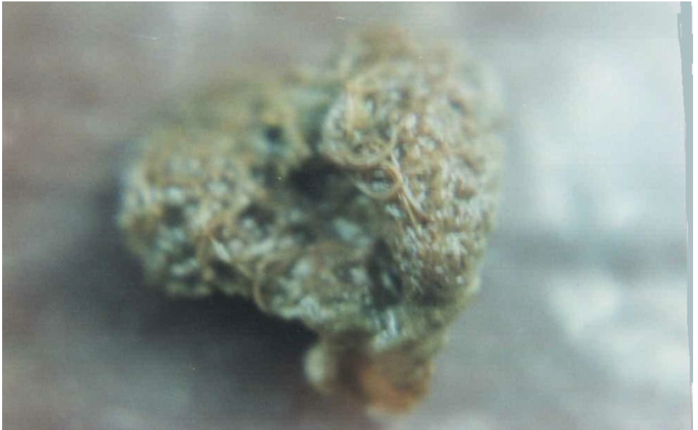
Macroscopically, the duodenum junction with the gizzard of both F. atra and G. chloropus was inflamed. Longitudinal cutting of this area showed a huge number of the nematode A. acutum (Fig. 1), blocking the lumen and preventing food passage. Small rough solid nodules were also observed on the intestinal wall of both birds.
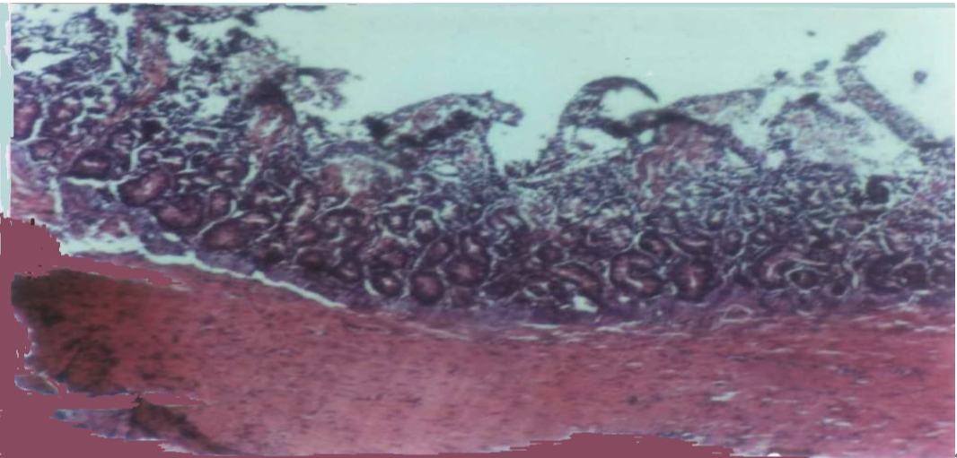
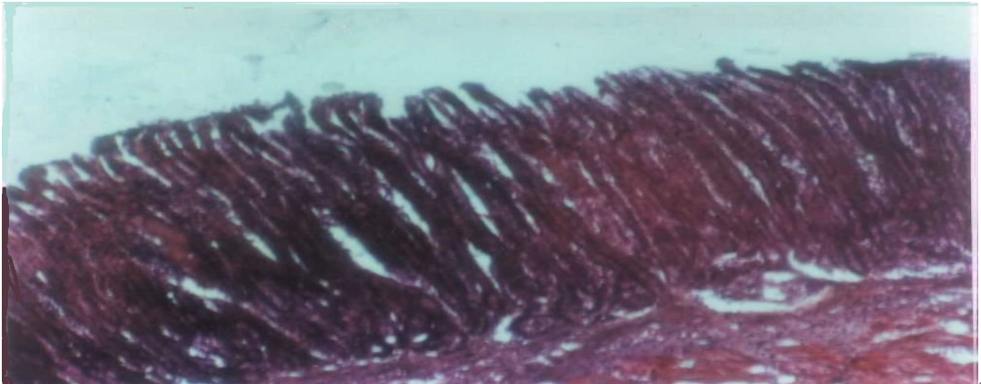
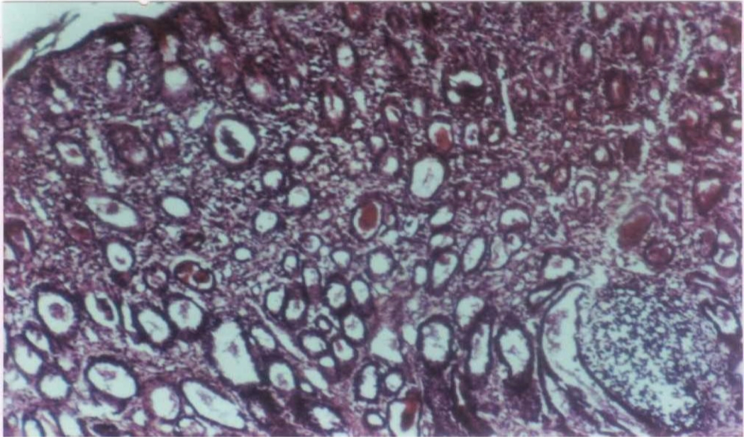
Microscopically, the most prominent feature was concerned mainly with villi because most of these structures showed atrophy, shortening, degeneration, and filled with inflammatory cells (Fig. 2) compared with their long and thin shape (Fig. 3).
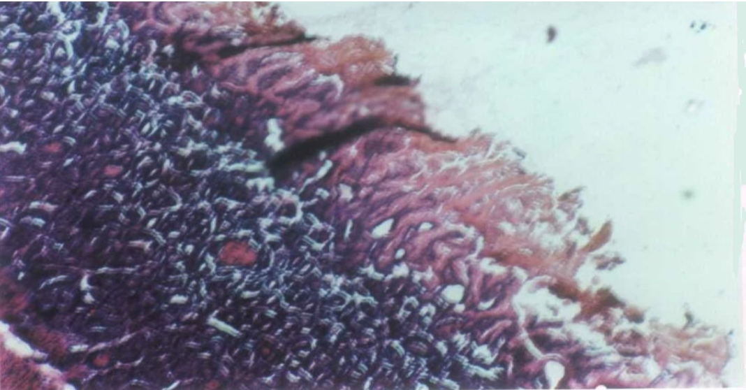
The degenerated villi appeared cracked, loosened, and lost their normal appearance (Fig. 4) compared to the normal villi (Fig. 3).
A large number of chronic inflammatory cells were noted, which filled the entire intestinal villi. Such cells also occurred in both the muscularis mucosa and serosa of the intestine of both bird species (Fig. 5).
An increase in the number of acini found in the base of the intestinal villi was also observed. Such acini appeared in a single row in the uninfected intestine (Fig. 2).
Discussion
A. acutum causes severe damage to its host by initiating anemia in addition to mechanical injuries to the host tissues as it tries to penetrate deeply and cannot be removed [13]. Among the mechanical damage observed in the present investigation was the occurrence of nodules on the internal walls.
Atrophy of villi occurred because of direct contact between the worms and the apex of villi, which causes a decrease in the villi absorption efficiency and a decrease in the absorption of the digested food, solutions, and necessary minerals [14,15].
In the intestine of both F. atra and G. chloropus infected with amidostomiasis in the present investigation, the villi lose their normal shape. Such degeneration was also observed in chickens infected with the cestode Raillietina cesticillus [16], domestic ducks infected with the trematode Notocotylus attenuatus [17], and in domestic fowl, Gallus gallus domesticus, suffering from cestodiasis [18].
Clear intestinal inflammation in the current study was represented by chronic inflammatory cells (lymphocytes, plasma cells, and phagocytes). A similar finding was also observed in domestic ducks infected with the nematode Echinuria uncinata [19]. Such lesions were observed as an increase in lymphocytes and eosinophils of domestic ducks infected with the trematode Sphaeridiotrema globulus [20]. An inflammatory response was noted in the proventriculus of the cattle egret Bubulcus ibis due to an infection with the nematode Microtetrameres egretes [21]. A similar response was also observed in the case of an infection of the little grebe P. ruficollis with the nematode E. tubifex [7]. The destruction of glandular secretory cells and a loss of secretory function of the proventriculus of the North Island kaka Nestor meridionalis septentrionalis were reported with a nematode Microtetrameres nestoris infestation [22]. The increase in the number of acini, which is found in the base of the intestinal villi, is caused by villi degeneration. Phalacrocorax carbo infestation with the cestode Haploparaxis crassirostris revealed the destruction of the villi epithelial lining and intestinal glands [23]. The present study confirmed the earlier results on the role of helminthic parasites on the health of aquatic birds.
 PDF Links
 PubReader
 ePub Link
 Full text via DOI
 Download Citation
 Print
Print


