


ņä£ļĪĀ
Disulfiram (DSF)ņØĆ 60ļģä ņØ┤ņāü ņĢīņĮöņś¼ņØśņĪ┤ņ”Ø ņ╣śļŻīņĀ£ļĪ£ ņé¼ņÜ®ļÉśņŚłļŗż[1]. ņĢīņĮöņś¼ ņäŁņĘ© Ēøä, ņŚÉĒāäņś¼ņØĆ alcohol dehydrogenaseņŚÉ ņØśĒĢ┤ acetaldehydeļĪ£ ņĀäĒÖśļÉśĻ│Ā, aldehyde dehydrogenase (ALDH)ņŚÉ ņØśĒĢ┤ acetateļĪ£ ļīĆņé¼ļÉ£ļŗż. DSFļŖö ALDHņØś ĒÜ©ņåī ĒÖ£ņä▒ņØä ņ¢ĄņĀ£ĒĢśņŚ¼ ņ▓┤ļé┤ņŚÉņä£ acetaldehydeļź╝ ņČĢņĀüņŗ£ĒéżĻ│Ā, ņØ┤ļŖö ļæÉĒåĄ, ņ¢┤ņ¦Ćļ¤¼ņøĆ, ĒÖŹņĪ░ ļō▒ņØś ļČłņŠīĒĢ£ ņ”ØņāüņØä ņ£Āļ░£ĒĢśņŚ¼ ņĢīņĮöņś¼ ņäŁņĘ©ļź╝ ņĀ£ĒĢ£ņŗ£Ēé©ļŗż[2]. ĒĢ£ĒÄĖ ņĄ£ĻĘ╝ņŚÉļŖö DSFļź╝ ĒĢŁņĢöņĀ£ļĪ£ ĒÖ£ņÜ®ĒĢśĻĖ░ ņ£äĒĢ£ ņŚ░ĻĄ¼Ļ░Ć ļŗżņ¢æĒĢ£ Ļ░üļÅäņŚÉņä£ ņ¦äĒ¢ēļÉśĻ│Ā ņ׳ļŗż[3].
DSFņØś ĒĢŁņĢöĒÜ©Ļ│╝ļŖö ņ£Āļ░®ņĢö[4-6], ĒÅÉņĢö[7], ĻĄÉļ¬©ņäĖĒżņóģ[8], ļīĆņןņĢö[6] ļō▒ņŚÉņä£ ņŚ░ĻĄ¼ļÉśņŚłņ£╝ļ®░, reactive oxygen species (ROS) ņāØņä▒[4], proteasome ĒÖ£ņä▒ ņ¢ĄņĀ£[5], nuclear factor-kappa B [6]ļéś mitogen-activated protein kinase [9]ņÖĆ Ļ░ÖņØĆ ņŗĀĒśĖņĀäļŗ¼ ņ▓┤Ļ│äļź╝ ņĪ░ņĀłĒĢśļŖö ļō▒ņØś ļŗżņ¢æĒĢ£ ĒĢŁņĢö ĻĖ░ņĀäņØ┤ ņĢīļĀżņĪīļŗż. ĒŖ╣Ē׳ copper (Cu)ņÖĆ Ļ▓░ĒĢ®ļÉśņŚłņØä ļĢīļŖö DSFņØś ĒĢŁņĢöĒÜ©Ļ│╝ļź╝ ņ”ØļīĆņŗ£ņ╝░ļŗż[6,8,9].
ļŗżņ¢æĒĢ£ ņĢöņŚÉņä£ DSFņØś ĒĢŁņĢöĒÜ©Ļ│╝Ļ░Ć ņŚ░ĻĄ¼ļÉśĻ│Ā ņ׳ļŖö Ļ░ĆņÜ┤ļŹ░, ļ”╝ĒöäņóģņŚÉ Ļ┤ĆĒĢ£ ņŚ░ĻĄ¼ļŖö ņĢäņ¦ü ļČĆņĪ▒ĒĢśļŗż. ļ│Ė ņŚ░ĻĄ¼ņŚÉņä£ļŖö, EL4 ņäĖĒżļź╝ ņØ┤ņÜ®ĒĢśņŚ¼ T ļ”╝ĒöäņóģņŚÉ ļīĆĒĢ£ DSFņØś ĒĢŁņĢöĒÜ©Ļ│╝ņÖĆ ņ×æņÜ®ĻĖ░ņĀäņØä ņĢīņĢäļ│┤ņĢśļŗż. ņØ┤ļź╝ ņ£äĒĢ┤, EL4 ņäĖĒżņŚÉ DSFļź╝ ļåŹļÅä ļ│äļĪ£ ņ▓śļ”¼ĒĢ£ Ēøä ņäĖĒżņØś ĒĢĄ ĒśĢĒā£ļź╝ Ļ┤Ćņ░░ĒĢśņśĆĻ│Ā, EL4 ņäĖĒżņØś ļīĆņé¼ĒÖ£ņä▒ļÅä, ļ»ĖĒåĀņĮśļō£ļ”¼ņĢä ļ¦ē ņĀäņ£ä(mitochondria membrane potential, MMP), ROS Ļ┤ĆļĀ©ņä▒ ļō▒ņØä ņĖĪņĀĢĒĢśņśĆļŗż. ļśÉĒĢ£, DSFĻ│╝ Cuļź╝ ĒĢ©Ļ╗ś ņ▓śļ”¼ĒĢśņŚ¼ CuĻ░Ć DSFņØś ĒÜ©ļŖźņŚÉ ļ»Ėņ╣śļŖö ņśüĒ¢źļÅä ņĢīņĢäļ│┤ņĢśļŗż.
ņ×¼ļŻī ļ░Å ļ░®ļ▓Ģ
ņäĖĒżņŻ╝ņÖĆ ņŗ£ņĢĮ
EL4 ņäĖĒżņŻ╝(mouse lymphoma cell line)ļŖö ĒĢ£ĻĄŁņäĖĒżņŻ╝ņØĆĒ¢ē(Korean Cell Line Bank)ņŚÉņä£ ĻĄ¼ņ×ģĒĢśņśĆļŗż. DSFļŖö Sigma-Aldrich (USA)ņŚÉņä£ ĻĄ¼ņ×ģĒĢśņśĆņ£╝ļ®░, dimethyl sulfoxide (DMSO; Sigma-Aldrich)ņŚÉ ļģ╣ņØĖ Ēøä ņé¼ņÜ®ĒĢśņśĆļŗż. DMSO ņé¼ņÜ®ļåŹļÅäļŖö DSF 10 ┬ĄMņØś Ļ▓ĮņÜ░ 1% (v/v)ļź╝ ņé¼ņÜ®ĒĢśņśĆņ£╝ļ®░, ļéśļ©Ėņ¦Ć ļ¬©ļōĀ DSF ņ▓śļ”¼ĻĄ░ņŚÉņä£ļŖö 0.1% (v/v) ņØ┤ĒĢśļĪ£ ņé¼ņÜ®ĒĢśņśĆļŗż. Cu2+ ņ▓śļ”¼ļź╝ ņ£äĒĢ┤ CuCl2 (Sigma-Aldrich)ļź╝ ļ®ĖĻĘĀ ņ”ØļźśņłśņŚÉ ļģ╣ņŚ¼ ņé¼ņÜ®ĒĢśņśĆļŗż.
ņäĖĒżļ░░ņ¢æĻ│╝ ļ¼╝ņ¦łņ▓śļ”¼
EL4 ņäĖĒżļŖö 10% fetal bovine serum, 100 U/mL penicillin/streptomycin, 2 mM L-glutamineņØä ĒżĒĢ©ĒĢ£ RPMI 1640 ļ░░ņ¦ĆņŚÉņä£ 37Ōäā, 5% CO2ņØś ņĪ░Ļ▒┤ņ£╝ļĪ£ ļ░░ņ¢æĒĢśņśĆļŗż. 6-, 24- ļśÉļŖö 96-well culture plate (ThermoFisher Scientific, USA)ņŚÉ ļČäņŻ╝ĒĢ£ ļÆż, DSFĻ│╝ CuCl2ļź╝ ļåŹļÅä ļ│äļĪ£ ņ▓śļ”¼ĒĢśņŚ¼ ļ░░ņ¢æĒĢ£ Ēøä ļČäņäØĒĢśņśĆļŗż.
EL4 ņäĖĒżņØś ļīĆņé¼ĒÖ£ņä▒ļÅä ņĖĪņĀĢ
1 ├Ś 105 cells/mL ļåŹļÅäņØś EL4 ņäĖĒżņŚÉ DSFļź╝ ļåŹļÅä ļ│äļĪ£ ņ▓śļ”¼ĒĢśĻ│Ā, CuCl2 1 ┬ĄMņØä ņČöĻ░ĆĒĢśņŚ¼, 96-well culture plateņŚÉņä£ 3ņØ╝ ļÅÖņĢł ļ░░ņ¢æĒĢśņśĆļŗż. ļ░░ņ¢æ Ēøä ņäĖĒżņØś ļīĆņé¼ĒÖ£ņä▒ļÅä ņĖĪņĀĢņØä ņ£äĒĢ┤ 3-(4,5-dimethylthiazol-2-yl)-2,5-diphenyltetrazoliumbromide (MTT, Sigma-Aldrich) ņÜ®ņĢĪņØä 0.5 mg/mL ļåŹļÅäļĪ£ ļäŻĻ│Ā 4ņŗ£Ļ░ä ļÅÖņĢł 37Ōäā, 5% CO2ņŚÉņä£ ņ▓śļ”¼ĒĢśņśĆļŗż. ņäĖĒżņŚÉ ņØśĒĢ┤ ĒÖśņøÉļÉ£ formazan product (crystal violet)ņØä ņÜ®ĒĢ┤ņŗ£ĒéżĻĖ░ ņ£äĒĢ┤ 10% sodium dodecyl sulfate ņÜ®ņĢĪņØä ļäŻņ¢┤ 2ņŗ£Ļ░ä ļÅÖņĢł ļ░śņØæņŗ£ņ╝░ļŗż. ĻĘĖ Ēøä microplate reader (ThermoFisher Scientific)ļź╝ ņØ┤ņÜ®ĒĢśņŚ¼ ĒØĪĻ┤æļÅä(570 nm)ļź╝ ņĖĪņĀĢĒĢśņśĆļŗż.
Hoechst 33342 ņŚ╝ņāēņØä ņØ┤ņÜ®ĒĢ£ EL4 ņäĖĒż ĒĢĄ Ļ┤Ćņ░░
ņäĖĒż ĒĢĄ ļ¬©ņ¢æņØś Ļ┤Ćņ░░ņØä ĒåĄĒĢ┤ ņäĖĒżņé¼ļź╝ ĒÖĢņØĖĒĢśĻĖ░ ņ£äĒĢ┤ EL4 ņäĖĒżņØś ĒĢĄņØä ņŚ╝ņāēĒĢśņśĆļŗż. 2 ├Ś 105 cells/mL ļåŹļÅäņØś EL4 ņäĖĒżņŚÉ DSFļź╝ ļåŹļÅä ļ│äļĪ£ ņ▓śļ”¼ĒĢśĻ│Ā CuCl2 1 ┬ĄMņØä ņČöĻ░ĆĒĢśņŚ¼, 24-well culture plateņŚÉņä£ 2ņØ╝ ļÅÖņĢł ļ░░ņ¢æĒĢśņśĆļŗż. ņäĖĒżņŚÉ Hoechst 33342 (Sigma-Aldrich) ņÜ®ņĢĪņØä 2.5 ╬╝g/mL ļåŹļÅäļĪ£ ņ▓śļ”¼ĒĢśņŚ¼ 37┬░CņŚÉņä£ 10ļČäĻ░ä ņŚ╝ņāēĒĢśņśĆļŗż. ĻĘĖ Ēøä ĒśĢĻ┤æĒśäļ»ĖĻ▓Į(ZOE Fluorescent Cell Imager; BIO-RAD, USA)ņØä ņØ┤ņÜ®ĒĢ┤ Ļ┤Ćņ░░ĒĢśņśĆļŗż.
ņäĖĒżņé¼ ņĖĪņĀĢ
ņäĖĒżņØś ņäĖĒżņé¼ ņĖĪņĀĢņØä ņ£äĒĢ┤ annexin V-fluorescein isothiocyanate (FITC)ņÖĆ propidium iodide (PI) ņÜ®ņĢĪņ£╝ļĪ£ ņŚ╝ņāēĒĢśņśĆļŗż. 2 ├Ś 105 cells/mL ļåŹļÅäņØś EL4 ņäĖĒżņŚÉ DSFļź╝ ļåŹļÅä ļ│äļĪ£ ņ▓śļ”¼ĒĢśĻ│Ā CuCl2 1 ┬ĄMņØä ņČöĻ░ĆĒĢśņŚ¼, 24-well culture plateņŚÉņä£ 2ņØ╝ ļÅÖņĢł ļ░░ņ¢æĒĢśņśĆļŗż. ļ░░ņ¢æņØ┤ ļüØļéśļ®┤ annexin V binding buffer (ThermoFisher Scientific)ļĪ£ ņäĖņ▓Ö Ēøä annexin V-FITC (BD Bioscience, USA)ļź╝ 2 ┬Ąg/mL ļåŹļÅäļĪ£ ņ▓śļ”¼ĒĢ£ ļŗżņØī ņāüņś©, ņĢöņŗżņØś ņĪ░Ļ▒┤ņŚÉņä£ 10ļČäĻ░ä ļ░śņØæņŗ£ņ╝░ļŗż. PI ņÜ®ņĢĪņØĆ ļČäņäØ 5ļČä ņĀä 0.1 ┬Ąg/mL ļåŹļÅäļĪ£ ņ▓śļ”¼ĒĢśņśĆļŗż. LSRFortessa flow cytometerņÖĆ FlowJo software (BD Bioscience)ļź╝ ņØ┤ņÜ®ĒĢśņŚ¼ ņ£ĀņäĖĒżļČäņäØĒĢśņśĆļŗż.
ROS Ļ┤ĆļĀ©ņä▒ ļČäņäØ
1 ├Ś 105 cells/mL ļåŹļÅäņØś EL4 ņäĖĒżņŚÉ DSFĻ│╝ 3 mM N-acetyl-L-cysteine (NAC, Sigma-Aldrich)ņØä ņ▓śļ”¼ĒĢ£ Ēøä 96-well culture plateņŚÉņä£ 3ņØ╝ ļÅÖņĢł ļ░░ņ¢æĒĢśņśĆļŗż. NACļŖö ROSņØś inhibitorļĪ£, ROSņØś Ļ┤ĆļĀ©ņä▒ ļČäņäØņØä ņ£äĒĢ┤ ņé¼ņÜ®ĒĢśņśĆļŗż. ļ░░ņ¢æ Ēøä ļīĆņé¼ĒÖ£ņä▒ļÅä ņĖĪņĀĢļ░®ļ▓Ģņ£╝ļĪ£ MTT assayļź╝ ņØ┤ņÜ®ĒĢ┤ņä£ ļČäņäØĒĢśņśĆļŗż. CuCl2Ļ░Ć ROSņØś ņāØņä▒ņŚÉ ļ»Ėņ╣śļŖö ņśüĒ¢źņØä ņĢīņĢäļ│┤ĻĖ░ ņ£äĒĢ┤ 2',7'-dichlorofluorescin diacetate (DCFDA; Sigma-Adrich)ļź╝ ņØ┤ņÜ®ĒĢśņŚ¼ ņ£ĀņäĖĒżļČäņäØņØä ĒĢśņśĆļŗż. DCFDAļŖö ROSņŚÉ ņØśĒĢ┤ 2',7' -dichlorofluorescin (DCF)ņ£╝ļĪ£ ņé░ĒÖöļÉśņ¢┤ ņĖĪņĀĢļÉ£ļŗż. 2 ├Ś 105 cells/mL ļåŹļÅäņØś EL4 ņäĖĒżņŚÉ DSFĻ│╝ CuCl2ļź╝ ņ▓śļ”¼ĒĢśņŚ¼ 2ņØ╝ ļÅÖņĢł ļ░░ņ¢æĒĢśņśĆļŗż. ļ░░ņ¢æņØ┤ ļüØļéśļ®┤ ņØĖņé░ņÖäņČ®ņĢĪņ£╝ļĪ£ ņäĖņ▓ÖĒĢ£ Ēøä DCFDAļź╝ 10 ┬ĄM ļåŹļÅäļĪ£ ņ▓śļ”¼ĒĢśņśĆļŗż. 37ŌäāņŚÉņä£ 30ļČäĻ░ä ļ░śņØæņŗ£Ēé© Ēøä ņ£ĀņäĖĒżļČäņäØ ņŚ╝ņāēņÜ®ņĢĪņ£╝ļĪ£ ņäĖņ▓ÖĒĢ£ ļÆż ļČäņäØĒĢśņśĆļŗż. ņ£ĀņäĖĒżļČäņäØņØĆ LSRFortessa flow cytometerņÖĆ FlowJo softwareļź╝ ņØ┤ņÜ®ĒĢśņśĆļŗż.
MMP ņĖĪņĀĢ
2 ├Ś 105 cells/mL ļåŹļÅäņØś EL4 ņäĖĒżņŚÉ DSFļź╝ ļåŹļÅä ļ│äļĪ£ ņ▓śļ”¼ĒĢśĻ│Ā CuCl2 1 ┬ĄMņØä ņČöĻ░ĆĒĢśņŚ¼, 6-well culture plateņŚÉņä£ 2ņØ╝ ļÅÖņĢł ļ░░ņ¢æĒĢśņśĆļŗż. MMP ņĖĪņĀĢņØä ņ£äĒĢ┤ Rhodamine 123 (Sigma-Aldrich) ņÜ®ņĢĪņØä 0.1 ┬Ąg/mL ļåŹļÅäļĪ£ ņ▓śļ”¼ĒĢ£ Ēøä ņāüņś©, ņĢöņŗżņØś ņĪ░Ļ▒┤ņŚÉņä£ 30ļČä Ļ░ä ļ░śņØæņŗ£ņ╝░ļŗż. LSRFortessa flow cytometerņÖĆ FlowJo softwareļź╝ ņØ┤ņÜ®ĒĢśņŚ¼ ņ£ĀņäĖĒżļČäņäØĒĢśņśĆļŗż.
ĒåĄĻ│äļČäņäØ
MTT assayņŚÉ ļīĆĒĢ£ Ļ▓░Ļ│╝ļŖö ĒÅēĻĘĀ ┬▒ Ēæ£ņżĆĒÄĖņ░©ļĪ£ ļéśĒāĆļāłņ£╝ļ®░, ordinary two-way ANOVA (GraphPad Prism; GraphPad Software, USA)ļź╝ ĒåĄĒĢ┤ ņ£ĀņØśņä▒ņØä ĒÖĢņØĖĒĢśņśĆļŗż. nsļŖö ņ£ĀņØśņä▒ņØ┤ ņŚåņØīņØä, *, **, ***, ****ļŖö Ļ░üĻ░ü ļīĆņĪ░ĻĄ░Ļ│╝ ļ╣äĻĄÉĒĢśņŚ¼ p < 0.05, 0.01, 0.001, 0.0001ņ×äņØä ļéśĒāĆļéĖļŗż. #ņØĆ Ļ░ÖņØĆ DSF ļåŹļÅäņŚÉņä£ NAC ņ▓śļ”¼ ņ£Āļ¼┤ņŚÉ ļö░ļźĖ ņ▓śļ”¼ĻĄ░ Ļ░äņØś pĻ░ÆņØä ļéśĒāĆļé┤ļ®░, Ļ░£ņłśņŚÉ ļö░ļźĖ ņ£ĀņØśņä▒ ņĀĢļÅäļŖö ņ£äņÖĆ ļÅÖņØ╝ĒĢśļŗż.
Ļ▓░Ļ│╝
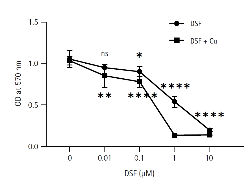
EL4 ņäĖĒżņŚÉ ļīĆĒĢ£ DSFņÖĆ CuCl2ņØś ļīĆņé¼ĒÖ£ņä▒ļÅä Ļ░Éņåī ĒÜ©Ļ│╝
DSF ļŗ©ļÅģ ļśÉļŖö DSF + CuCl2 (1 ╬╝M) ņ▓śļ”¼ĻĄ░ņŚÉņä£ EL4 ņäĖĒżņØś ļīĆņé¼ĒÖ£ņä▒ļÅä ļ│ĆĒÖöļź╝ ņĢīņĢäļ│┤ĻĖ░ ņ£äĒĢ┤ MTT assayļź╝ ņŗ£Ē¢ēĒĢśņśĆļŗż(Fig. 1). ļČäņäØ Ļ▓░Ļ│╝ DSFņØś ļ¬©ļōĀ ļåŹļÅä(0.01-10 ┬ĄM)ņŚÉņä£ DSF ļŗ©ļÅģņŚÉ ļ╣äĒĢ┤ DSF + CuCl2 ņ▓śļ”¼ĻĄ░ņØ┤ ļé«ņØĆ ļīĆņé¼ĒÖ£ņä▒ļÅäļź╝ ļéśĒāĆļāłļŗż. ĒŖ╣Ē׳ DSF 1 ┬ĄMņŚÉņä£ ļīĆņĪ░ĻĄ░Ļ│╝ ļ╣äĻĄÉĒ¢łņØä ļĢī DSF ļŗ©ļÅģņØĆ 49%, DSF + CuCl2 ņ▓śļ”¼ĻĄ░ņØĆ 87%ņØś ļīĆņé¼ĒÖ£ņä▒ļÅä Ļ░Éņåīļź╝ ļ│┤ņśĆļŗż. DSF ļŗ©ļÅģņ▓śļ”¼ĻĄ░ņØś Ļ▓ĮņÜ░ 0.01 ┬ĄMņŚÉņä£ ļīĆņé¼ĒÖ£ņä▒ļÅäĻ░Ć ņĢĮĻ░ä Ļ░ÉņåīĒĢśņśĆņ¦Ćļ¦ī ņ£ĀņØśĒĢśņ¦Ć ņĢŖņĢśĻ│Ā, 10 ┬ĄMņŚÉņä£ļŖö ļīĆņé¼ĒÖ£ņä▒ņØ┤ Ļ▒░ņØś ļ│┤ņØ┤ņ¦Ć ņĢŖņĢśļŗż. ņØ┤ļŖö DSFĻ░Ć 0.01 ┬ĄM ņØ┤ĒĢśņØś ļåŹļÅäņŚÉņä£ļŖö sub-cytotoxicĒĢśļŗżļŖö Ļ▓āņØä ļéśĒāĆļéĖļŗż. Half maximal inhibitory concentration (IC50)ņØä Ļ│äņé░ĒĢ£ Ļ▓░Ļ│╝ DSF ļŗ©ļÅģņØĆ 1.02 ┬ĄM, DSF + CuCl2 ņ▓śļ”¼ĻĄ░ņØĆ 0.25 ┬ĄMņ£╝ļĪ£, DSF ļŗ©ļÅģņŚÉ ļ╣äĒĢ┤ CuCl2ņÖĆ ĒĢ©Ļ╗ś ņ▓śļ”¼Ē¢łņØä ļĢī ņäĖĒżļÅģņä▒ņØ┤ ņĢĮ 4ļ░░ ņ”ØĻ░ĆĒĢśņśĆņØīņØä ņĢī ņłś ņ׳ņŚłļŗż.
DSFņÖĆ CuCl2ņŚÉ ņØśĒĢ£ ņäĖĒżņé¼ ņ”ØĻ░Ć
ņäĖĒżĒĢĄņØś ĒśĢĒā£ĒĢÖņĀü Ļ┤Ćņ░░ņØä ĒåĄĒĢ┤ ņäĖĒżņ×Éļ®Ėņé¼Ļ░Ć ņØ╝ņ¢┤ļéśļŖöņ¦Ć ĒÖĢņØĖĒĢśĻĖ░ ņ£äĒĢ┤ Hoechst 33342Ļ│╝ PIļĪ£ ņäĖĒżļź╝ ņŚ╝ņāēĒĢśņśĆļŗż(Fig. 2A). ņé┤ņĢäņ׳ļŖö ņäĖĒż(Ēīīļ×Ćņāē)ņÖĆ ņŻĮņØĆ ņäĖĒż(ļČēņØĆņāēĻ│╝ ļČäĒÖŹņāē)ļź╝ ņāēĻ╣öļĪ£ ĻĄ¼ļČäĒĢśņŚ¼ ļČäņäØĒĢśņśĆļŗż. DSF ļŗ©ļÅģņ▓śļ”¼ĻĄ░ņØĆ ļīĆņĪ░ĻĄ░Ļ│╝ ņŻĮņØĆ ņäĖĒżņØś ņłśĻ░Ć ļ╣äņŖĘĒĢśņśĆĻ│Ā, CuCl2ņÖĆ ĒĢ©Ļ╗ś ņ▓śļ”¼Ē¢łņØä ļĢīļŖö DSF 0.1 ┬ĄM + CuCl2ņŚÉņä£ ĒśäņĀĆĒĢśĻ▓ī ņŻĮņØĆ ņäĖĒżņØś ļ╣äņ£©ņØ┤ ļåÆņĢśļŗż(Fig. 2B). ļśÉĒĢ£ annexin V-FITC/PI ņŚ╝ņāē Ēøä ņ£ĀņäĖĒżļČäņäØņØä ņŗżņŗ£ĒĢśņŚ¼ ņäĖĒżņé¼ļź╝ ļŗ©Ļ│äļ│äļĪ£ ļČäņäØĒĢśņśĆļŗż(Fig. 2C). Necrosis (annexin V-/PI+), early apoptosis (annexin V+/PI-), late apoptosis (annexin V+/PI+)ļĪ£ ĻĄ¼ļČäĒĢśņśĆļŗż. DSF 0.1 ┬ĄMņŚÉ ļ╣äĒĢ┤ DSF 0.1 ┬ĄM + CuCl2ņŚÉņä£ early apoptosisņÖĆ late apoptosisĻ░Ć ļŹö ņ”ØĻ░ĆĒ¢łļŗż. ĒŖ╣Ē׳ DSF 0.1 ┬ĄM + CuCl2ņ▓śļ”¼ĻĄ░ņØĆ ņĀä ņŗżĒŚśĻĄ░ņŚÉņä£ viable cell (annexin V-/PI-)ņØś ļ╣äņ£©ņØ┤ Ļ░Ćņן ļé«ņĢśļŗż. ņØ┤ļź╝ ĒåĄĒĢ┤ DSFļŖö CuCl2ņŚÉ ņØśĒĢ┤ EL4 ņäĖĒżņØś ņäĖĒżņé¼ļź╝ ļŹöņÜ▒ ņ”Øņ¦äņŗ£Ēé¼ ņłś ņ׳ņØīņØä ņĢī ņłś ņ׳ņŚłļŗż.
DSFņŚÉ ņØśĒĢ£ EL4 ņäĖĒżņØś MMP Ļ░Éņåī
MMPļŖö ļ»ĖĒåĀņĮśļō£ļ”¼ņĢäņØś ĻĖ░ļŖźņØä ņ£Āņ¦ĆĒĢśĻĖ░ ņ£äĒĢ£ ĒĢäņłśņĪ░Ļ▒┤ņØ┤ļŗż. ņ£ĀņäĖĒżļČäņäØņŚÉņä£ forward scatter/side scatterņŚÉ ĻĘ╝Ļ▒░ĒĢśņŚ¼ debrisļĪ£ ņČöņĀĢļÉśĻ▒░ļéś ņŻĮņØĆ ņäĖĒżļŖö ļ░░ņĀ£ĒĢ£ Ēøä ņé┤ņĢäņ׳ļŖö ņäĖĒżļōżņØä gatingĒĢśņŚ¼ MMP Ļ░ÆņØä ņĖĪņĀĢĒ¢łļŗż(Fig. 3). DSF 0.1 ╬╝M + CuCl2 (1 ╬╝M)ņŚÉņä£ fluorescence intensity ĒÅēĻĘĀĻ░ÆņØĆ Ļ░Ćņן ĒśäņĀĆĒ׳ ļé«ņĢśļŖöļŹ░, ļīĆņĪ░ĻĄ░ņŚÉ ļ╣äĒĢ┤ 48% ļé«ņĢśļŗż. DSF 0.02 ╬╝M ļåŹļÅäņÖĆ 0.1 ╬╝M ļåŹļÅä ļ¬©ļæÉņŚÉņä£ CuCl2 (1 ╬╝M)ļź╝ ĒĢ©Ļ╗ś ņ▓śļ”¼Ē¢łņØä ļĢīĻ░Ć DSF ļŗ©ļÅģņ▓śļ”¼ĻĄ░ļ│┤ļŗż ĒÅēĻĘĀĻ░ÆņØ┤ 40% ļé«ņĢśļŗż. DSFļź╝ CuCl2ņÖĆ ĒĢ©Ļ╗ś ņé¼ņÜ®ĒĢĀ Ļ▓ĮņÜ░ EL4 ņäĖĒżņØś ļ»ĖĒåĀņĮśļō£ļ”¼ņĢä ĻĖ░ļŖźņØä ĒÜ©Ļ│╝ņĀüņ£╝ļĪ£ ņ¢ĄņĀ£ĒĢĀ ņłś ņ׳ņØīņØä ĒÖĢņØĖĒ¢łļŗż.
DSFņŚÉ ņØśĒĢ£ ĒĢŁņĢöĒÜ©Ļ│╝ņŚÉņä£ ROSņØś Ļ┤ĆļĀ©ņä▒
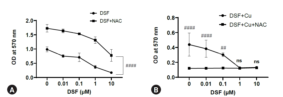
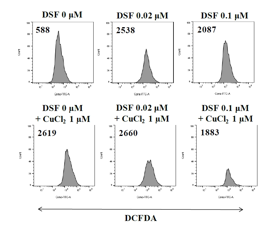
DSFņØś ĒĢŁņĢöĒÜ©Ļ│╝ņŚÉņä£ ROSĻ░Ć ņ¢┤ļ¢╗Ļ▓ī Ļ┤ĆļĀ©ļÉśļŖöņ¦Ć ņĢīņĢäļ│┤ĻĖ░ ņ£äĒĢ┤ ROSņØś inhibitor ņżæ ĒĢśļéśņØĖ NACļź╝ ņé¼ņÜ®ĒĢśņśĆļŗż. DSFņØś ņ▓śļ”¼ ļåŹļÅäļ▓öņ£ä(0-10 ┬ĄM)ņŚÉņä£ DSF ļŗ©ļÅģņ▓śļ”¼ĻĄ░ļ│┤ļŗż DSFņÖĆ NACļź╝ ĒĢ©Ļ╗ś ņ▓śļ”¼Ē¢łņØä ļĢī ņäĖĒżņØś ļīĆņé¼ĒÖ£ņä▒ļÅäĻ░Ć ļåÆņØĆ Ļ▓āņ£╝ļĪ£ ļ│┤ņĢä, ROSĻ░Ć DSFļĪ£ ņ▓śļ”¼ļÉ£ EL4 ņäĖĒżņØś ļīĆņé¼ĒÖ£ņä▒ļÅäļź╝ ļé«ņČöļŖö ņé¼ņŗżņØä ņĢī ņłś ņ׳ņŚłļŗż(Fig. 4A). ĒĢśņ¦Ćļ¦ī DSF + CuCl2 (1 ╬╝M) ņ▓śļ”¼ĻĄ░ņŚÉ NACļź╝ ļŹöĒĢ┤ ļ░śņØæņŗ£Ēéżļ®┤ DSFņØś ļ¬©ļōĀ ļåŹļÅäņŚÉņä£ ņäĖĒżņØś ļīĆņé¼ĒÖ£ņä▒ļÅäĻ░Ć ļ¦żņÜ░ ļ¢©ņ¢┤ņĪīļŗż(Fig. 4B). DSF + CuCl2 ņ▓śļ”¼ĻĄ░ņØś ROS ņāØņä▒ļ¤ēņØä ņĢīņĢäļ│┤ĻĖ░ ņ£äĒĢ┤ DCFDAļź╝ ņØ┤ņÜ®ĒĢśņśĆļŗż. DSF ļśÉļŖö CuCl2 ņ▓śļ”¼ĻĄ░ņØĆ ļīĆņĪ░ĻĄ░ņŚÉ ļ╣äĒĢ┤ ROS ņāØņä▒ļ¤ēņØ┤ ņ”ØĻ░ĆĒ¢łļŗż(Fig. 5).
Ļ│Āņ░░
DSFļŖö 60ļģä ņØ┤ņāü ņĢīņĮöņś¼ņØśņĪ┤ņ”Ø ņ╣śļŻīņĀ£ļĪ£ ņé¼ņÜ®ļÉśņ¢┤ ņÖöļŗż[1]. ņØ┤Ēøä DSFĻ░Ć ņĢīņĮöņś¼ņØśņĪ┤ņ”Ø ņ╣śļŻīņĀ£ļĪ£ ņé¼ņÜ®ļÉśļŖö ĻĖ░ņĀäĻ│╝ļŖö ļŗżļźĖ ĻĖ░ņĀäņ£╝ļĪ£ ĒĢŁņĢöņ×æņÜ®ņØä ĒĢ£ļŗżļŖö ņŚ░ĻĄ¼ļÅä ņ¦äĒ¢ēļÉśņŚłļŗż[4-6]. ļśÉĒĢ£ ļīĆĻĘ£ļ¬©ļĪ£ 4,910ņóģņØś ņĢĮļ¼╝ ļ░Å Ēøäļ│┤ļ¼╝ņ¦łņØä ņŗżĒŚśĒĢ£ Ļ▓░Ļ│╝ DSFĻ░Ć ņĀäļ”ĮņäĀ ņĢöņäĖĒżņØś ņä▒ņןņØä ņ¢ĄņĀ£ĒĢśļŖö ļ¼╝ņ¦łļĪ£ ļ░ØĒśĆņ¦ĆĻĖ░ļÅä Ē¢łļŗż[10]. DSFļŖö ļ╣äĻĄÉņĀü Ļ░ĆĻ▓®ņØ┤ ņĀĆļĀ┤ĒĢśĻ│Ā, ņé¼ļ×īņŚÉņä£ ļČĆņ×æņÜ®Ļ│╝ ļÅģņä▒ņŚÉ Ļ┤ĆĒĢ£ ņŚ░ĻĄ¼Ļ░Ć ņČ®ļČäĒ׳ ņ¦äĒ¢ēļÉśņŚłĻĖ░ ļĢīļ¼ĖņŚÉ ĒĢŁņĢöņĀ£ļĪ£ņŹ© repurposing drugņ£╝ļĪ£ ņé¼ņÜ®ļÉĀ ņłś ņ׳ļŖö Ļ░ĆļŖźņä▒ņØä ņ¦Ćļģöļŗż. ņØ┤ļ¤¼ĒĢ£ DSFņØś ĒĢŁņĢöĒÜ©Ļ│╝ļŖö CuņÖĆ ĒĢ©Ļ╗ś ņ▓śļ”¼ļÉÉņØä ļĢī ļŹöņÜ▒ ņ”ØĻ░ĢļÉśļ®░, ļŗżņ¢æĒĢ£ ņĢöņŚÉņä£ DSFņÖĆ CuņØś ĒĢŁņĢöĒÜ©Ļ│╝ņŚÉ ļīĆĒĢ£ ņŚ░ĻĄ¼Ļ░Ć ņØ┤ļŻ©ņ¢┤ņĪīļŗż[8,9]. ļ│Ė ņŚ░ĻĄ¼ņŚÉņä£ļŖö ņĢģņä▒ T ļ”╝ĒöäņóģņŚÉņä£ ņØ┤ļź╝ ņŚ░ĻĄ¼ĒĢśņśĆļŗż.
DSFņØś ļ¬©ļōĀ ļåŹļÅä(0.01-10 ┬ĄM)ņŚÉņä£ DSF ļŗ©ļÅģņŚÉ ļ╣äĒĢ┤ DSF + CuCl2 ņ▓śļ”¼ĻĄ░ņØ┤ ļŹö ļé«ņØĆ ļīĆņé¼ĒÖ£ņä▒ļÅäļź╝ ļ│┤ņśĆļŗż(Fig. 1). CuCl2ņØś ņ£Āļ¼┤ņŚÉ ļö░ļźĖ ņ░©ņØ┤ļŖö DSF 1 ┬ĄMņŚÉņä£ Ļ░Ćņן ņ╗Ėļŗż. DSF + CuCl2ņØś ņäĖĒżĒÖ£ņä▒ļÅä ņĀĆĒĢśņŚÉ ļīĆĒĢ£ ņ×æņÜ®ĻĖ░ņĀäņØä ļČäņäØĒĢśĻĖ░ ņ£äĒĢ┤ ņäĖĒżņé¼ ņŚ¼ļČĆ, ļ»ĖĒåĀņĮśļō£ļ”¼ņĢä ĻĖ░ļŖź ļō▒ņØä ļČäņäØĒĢśņśĆļŗż. Hoechst 33342ļŖö Ļ▒┤Ļ░ĢĒĢ£ ņäĖĒżļ¦ēņØä Ļ┤ĆĒåĄĒĢśņŚ¼ DNAļź╝ Ēīīļ×Ćņāēņ£╝ļĪ£ ņŚ╝ņāēĒĢ£ļŗż[11]. ļ░śļ®┤ PIļŖö Ļ▒┤Ļ░ĢĒĢ£ ņäĖĒżļ¦ēņØä ĒåĄĻ│╝ĒĢśņ¦Ć ļ¬╗ĒĢśĻ│Ā ņåÉņāüļÉ£ ņäĖĒżņØś ņŚ╝ņāēņ▓┤ļź╝ ļ╣©Ļ░Ģņāēņ£╝ļĪ£ ņŚ╝ņāēĒĢ£ļŗż. Hoechst 33342/PI ņŚ╝ņāēņØä ĒåĄĒĢ┤ DSFņÖĆ CuCl2ļź╝ ĒĢ©Ļ╗ś ņ▓śļ”¼Ē¢łņØä ļĢī ņäĖĒżņé¼Ļ░Ć Ļ░Ćņן ņ”ØĻ░ĆĒ¢łļŗż(Fig. 2). ļśÉĒĢ£ Annexin V/PI ņŚ╝ņāēņØä ĒåĄĒĢ┤ ņäĖĒżņé¼ļź╝ ļŗ©Ļ│äņĀüņ£╝ļĪ£ ļéśļłäņ¢┤ ņłśņ╣śĒÖö Ē¢łņ£╝ļ®░, DSF 0.1 ┬ĄM + CuCl2ņŚÉņä£ ņé┤ņĢäņ׳ļŖö ņäĖĒż ņłśĻ░Ć Ļ░Ćņן ļé«ņØĆ ņé¼ņŗżņØä ĒÖĢņØĖĒ¢łļŗż.
ņäĖĒżņé¼ņØś ĻĖ░ņĀäņ£╝ļĪ£ MMPņÖĆ ROSņØś Ļ┤ĆļĀ©ņä▒ņØä ņĢīņĢäļ│┤ņĢśļŗż. Rhodamine 123ņØĆ ļ»ĖĒåĀņĮśļō£ļ”¼ņĢäņØś ņäĖĒżļ¦ēņØä ĒåĄĻ│╝ĒĢśļŖö ĒśĢĻ┤æņŚ╝ļŻīļĪ£, ņĀĢņāüņäĖĒżņŚÉņä£ļŖö ļ»ĖĒåĀņĮśļō£ļ”¼ņĢä ļé┤ņŚÉ ņČĢņĀüļÉśņ¦Ćļ¦ī, ņåÉņāüļÉ£ MMPļź╝ Ļ░Ćņ¦ä ņäĖĒżņŚÉņä£ļŖö ņČĢņĀüļÉśņ¦Ć ņĢŖļŖöļŗż[12]. ROSļŖö ļ»ĖĒåĀņĮśļō£ļ”¼ņĢäņØś ņĀäņ×ÉņĀäļŗ¼Ļ│äņŚÉņä£ ņĀĢņāüņĀüņ£╝ļĪ£ ņāØņä▒ļÉśļŖö Ļ│╝ņé░ĒÖöļ¼╝(peroxide) ļō▒ņØä ļ¦ÉĒĢśļ®░, ņ”ØĻ░ĆļÉ£ ROSļŖö ņäĖĒżņ×Éļ®Ėņé¼ ņĪ░ņĀł ļŗ©ļ░▒ņ¦ł(apoptosis regulatory protein)ņØś ļ░£ĒśäņŚÉ Ļ┤ĆņŚ¼ĒĢśņŚ¼ ņäĖĒżņé¼ļź╝ ņØ╝ņ£╝Ēé©ļŗż[13]. ļæÉ Ļ░Ćņ¦Ć ĻĖ░ņĀäņŚ░ĻĄ¼ļź╝ ĒåĄĒĢ┤ DSFĻ░Ć MMPņØś Ļ░ÉņåīņÖĆ ROSņØś ņāØņä▒ņØä ņ£ĀļÅäĒĢśņśĆĻ│Ā, CuCl2ļź╝ ĒĢ©Ļ╗ś ņ▓śļ”¼Ē¢łņØä ļĢī ņäĖĒżņé¼Ļ░Ć ņ”ØĻ░ĆĒĢ£ Ļ▓āņ£╝ļĪ£ ļ│┤ņĢä ņāüņŖ╣ĒÜ©Ļ│╝Ļ░Ć ņ׳ļŗżļŖö ņé¼ņŗżņØä ņĢī ņłś ņ׳ņŚłļŗż. ļśÉĒĢ£ NACļź╝ ņØ┤ņÜ®ĒĢ£ ROS ņāØņä▒ļ¤ē ļČäņäØņŚÉņä£ DSF + CuCl2 ņ▓śļ”¼ĻĄ░ņØś ļīĆņé¼ĒÖ£ņä▒ļÅäĻ░Ć DSFņØś ļ¬©ļōĀ ļåŹļÅäņŚÉņä£ ļ¦żņÜ░ ļ¢©ņ¢┤ņĪīļŖöļŹ░, ņØ┤ļŖö CuCl2 + NACĻ░Ć ņäĖĒżļÅģņä▒ ņ×æņÜ®ņØä ļéśĒāĆļéĖ Ļ▓āņ£╝ļĪ£ ĒīÉļŗ©ļÉ£ļŗż[14]. ļśÉĒĢ£ Ļ│©ņłśņóģņäĖĒżņŚÉņä£ DSF ļŗ©ļÅģņŚÉ ļ╣äĒĢ┤ DSFņÖĆ Cuļź╝ ĒĢ©Ļ╗ś ņé¼ņÜ®Ē¢łņØä ļĢī JNK ņŗĀĒśĖņĀäļŗ¼ņØ┤ ĒÖ£ņä▒ĒÖöļÉśņŚłļŗż[9]. Ē¢źĒøä EL4 ņäĖĒżņŚÉņä£ļÅä CuņØś ņāüņŖ╣ĒÜ©Ļ│╝ņÖĆ Ļ┤ĆļĀ©ļÉ£ ņŗĀĒśĖņĀäļŗ¼ņŚÉ ļīĆĒĢ£ ņŚ░ĻĄ¼Ļ░Ć ĒĢäņÜöĒĢśļŗż.
ļ│Ė ņŚ░ĻĄ¼ņŚÉņä£ DSF + CuņØś ĒĢŁņĢöĒÜ©Ļ│╝ļź╝ ĒÖĢņØĖĒ¢łņØīņŚÉļÅä, DSFĻ░Ć ĒĢŁņĢöņĀ£ļĪ£ Ļ░£ļ░£ļÉśļŖö ļŹ░ņŚÉļŖö ļ¬ć Ļ░Ćņ¦Ć ļŗ©ņĀÉņØ┤ ņ׳ļŗż. DSFļŖö ĒśłņĢĪ(pH 7.4) ļé┤ņŚÉņä£ ļ░śĻ░ÉĻĖ░Ļ░Ć 1-1.5ļČäņ£╝ļĪ£ ļ¦żņÜ░ ņ¦¦ņĢä[15], ĒśłņĢĪņŚÉņä£ ļīĆņé¼ļÉśļŖö Ļ│╝ņĀĢņŚÉņä£ ĒĢŁņĢöļŖźļĀźņØ┤ ļ¢©ņ¢┤ņ¦ł ņłś ņ׳ļŗż. ļŗżņ¢æĒĢ£ ņĢöņŚÉņä£ ĒĢŁņĢöņĀ£Ļ░Ć Ēæ£ņĀüņןĻĖ░ņŚÉ ĒÜ©Ļ│╝ņĀüņ£╝ļĪ£ ņ╣©Ēł¼ĒĢśĻ│Ā ņśżļל ļ©Ėļ¼╝Ļ▓ī ĒĢśĻĖ░ ņ£äĒĢ┤ nanoparticleļĪ£ packagingĒĢśņŚ¼ ņāØņ▓┤ņŚÉ ņĀüņÜ®ĒĢśļŖö ļ░®ļ▓ĢņØ┤ ņŚ░ĻĄ¼ļÉśĻ│Ā ņ׳ļŗż[16,17]. ļö░ļØ╝ņä£ DSFļÅä ļŗ©ļÅģ ļśÉļŖö DSF + Cuļź╝ ņāłļĪ£ņÜ┤ drug delivery system (DDS)ņ£╝ļĪ£ ņĀæĻĘ╝ĒĢĀ ĒĢäņÜöĻ░Ć ņ׳ļŗż. ņØ┤ļ▓ł ņŚ░ĻĄ¼ņŚÉņä£ DSFņØś T ļ”╝ĒöäņóģņŚÉ Ļ┤ĆĒĢ£ ĒĢŁņĢöĒÜ©Ļ│╝Ļ░Ć in vitroņŚÉņä£ ĒÖĢņØĖļÉśņŚłņ£╝ļ»ĆļĪ£, Ē¢źĒøä DSFņØś in vivoņŚÉņä£ ĒĢŁņĢöĒÜ©Ļ│╝ņŚÉ ļīĆĒĢ£ ņŚ░ĻĄ¼ņÖĆ ļ”╝ĒöäņóģņŚÉ DSF ļśÉļŖö DSF + Cuļź╝ ĒÜ©Ļ│╝ņĀüņ£╝ļĪ£ ņĀäļŗ¼ĒĢĀ ņłś ņ׳ļŖö DDS ņŚ░ĻĄ¼Ļ░Ć ĒĢäņÜöĒĢśļŗż.


 PDF Links
 PubReader
 ePub Link
 Full text via DOI
 Download Citation
 Print
Print


