
Introduction
Canine atopic dermatitis (AD) refers to genetically predisposed allergic dermatosis that causes chronic pruritus associated with exposure to numerous types of environmental allergens [1]. It is generally associated with typical clinical signs and the production of allergen-specific immunoglobulin E (IgE) antibodies [2]. In past decades, the prevalence of canine AD has increased [3]. Various strategies have been proposed to alleviate the clinical signs of canine AD including avoidance of allergens, application of anti-inflammatory drugs, and allergen-specific immunotherapy [4]. Allergen-specific immunotherapy is not a common treatment for canine AD because it requires the long-term subcutaneous or sublingual administration of allergens [4]. On the other hand, administration of anti-inflammatory and anti-pruritus drugs including topical/systemic glucocorticoids, ciclosporin, and oclacitinib is the main treatment option for canine AD. However, since long-term or high-dose use of these drugs has been associated with some adverse effects, concomitantly, a variety of adjunctive methods in the treatment of canine AD have been attempted to reduce the dosage of these drugs.
Studies have reported that probiotics, which are one of the adjunctive treatments and include specific microorganisms, could be effective against canine AD [5-7]. The effect of bacteria that produce lactate on canine AD has been described in the International Committee on Allergic Diseases of Animals (ICADA) guidelines [4]. Likewise, numerous human studies using different strains of probiotics have reported probiotics to be effective in improving the clinical signs of AD in young children [8-10]. Although the exact mechanism of the immunomodulatory effect of probiotics in human and canine AD has not been fully understood, a close relationship between intestinal bacterial flora and the numerous factors associated with the pathogenesis of AD has been suggested [11].
The main probiotics used for the treatment of human atopic patients are mainly Lactobacilli and Bifidobacteria. This is because the number of Lactobacilli and Bifidobacteria is significantly lower in the fecal samples of human infants and children with AD than in healthy individuals [12]. A study of human AD reported that pruritus and medication scores were improved by the administration of probiotics consisting of Lactobacilli strain [13]. Another study also reported great improvements in the clinical signs in atopic patients after combined administration of Bifidobacteria stain [10]. However, there have been few studies on the therapeutic or complementary effects of probiotics, especially Bifidobacterium strain, in canine AD. Therefore, we aimed to evaluate the beneficial effects of oral administration of Bifidobacterium (B.) longum in dogs with AD.
Materials and Methods
Animals
We enrolled 11 client-owned dogs that had been diagnosed with mild to moderate AD and treated according to ICADA guidelines [4] at the Veterinary Teaching Hospital of Chungbuk National University (Cheongju, South Korea). The diagnostic criteria for canine AD were based on the medical history, typical clinical signs according to the ICADA guidelines [4], fulfillment of more than 5 postulates in Favrot's criteria [14], and/or a positive intradermal skin test. To rule out cutaneous adverse food reactions, we performed a diet elimination trial for at least 8 weeks using a commercial hydrolyzed hypoallergenic diet, Royal Canin Hypoallergenic and Anallergenic (Royal Canin; Aimargues, France). We ruled out the possibility of other diseases that can cause pruritus and skin lesions through proper tests and treatments. The inclusion criteria included: a minimum score of 3 in the pruritus visual analog scale (PVAS) or of 30 in the Canine Atopic Dermatitis Extent and Severity Index (CADESI)-4 [15,16].
The dogs were randomly allocated to 2 groups; namely, the probiotics and control group. The probiotics group (n = 7) was prescribed with B. longum while the control group (n = 4) received a placebo powder. All formerly prescribed drugs for controlling canine AD were continued during the study period. We obtained informed consent from all the owners prior to the experiment.
Administration of probiotics
We obtained B. longum from the National Institute of Animal Science, Rural Development Administration. It had been isolated from feces of healthy Korean neonates and anaerobically cultured (5 × 10¹⁰ colony-forming units per 2 g). Probiotics in sachets were stored at 4°C and orally administered to the dog once a day for 12 consecutive weeks.
Assessments
Assessment of the CADESI-4 score, transepidermal water loss (TEWL), and medication score was conducted by veterinarians while that of PVAS was performed by the dog-owners. All clinical indices were surveyed every 4 weeks starting from the baseline to the 12 weeks.
PVAS
The severity of pruritus was graded using the PVAS score [16]. The PVAS values were determined based on history-taking and evaluation by the owners. The PVAS consists of 10 scales with 0 indicating no pruritus and 10 indicating the most severe pruritus.
CADESI-4
The extent and severity of skin lesions were evaluated using the CADESI-4, which was designed to assess 20 different body parts. We assessed 3 skin lesions including erythema, lichenification, and excoriation/alopecia based on a four-point severity scale. The scale indicated none (score 0), mild (score 1), moderate (score 2), or severe (score 3). We assessed 20 body sites, 3 types of lesion, and 4 grades of severity, and thus generating a maximal score of 20 × 3 × 3 = 180. The CADESI-4 cut-off scores for mild, moderate, and severe AD were 10, 35, and 60, respectively [15].
TEWL
Prior to assessing TEWL, the dogs were allowed to acclimate to the environment of the test room. We measured TEWL using an unventilated closed-chamber device, VaporMeter SWL3 (Delfin Technologies Ltd., Kuopio, Finland) according to the manufacturer's instructions. To minimize the TEWL variability, we performed five consecutive measurements for 10 seconds at a time on both pinnae, axillae, and the groin in a constant environment (ambient temperature: 24-25°C; relative humidity: 44-66%). Subsequently, we excluded the maximum and minimum values and averaged the remaining results and presented the average as g/m2/h.
Medication score
Medication score used in this study was based on previously described [7] and we used modified version. Alteration of consumed doses of the drugs including prednisolone, oclacitinib, and ciclosporin was calculated and converted into a medication score. A medication score of 1.0 was assigned to the drug dose that was being administered prior to the start of probiotics administration.
Statistical analysis
Data were analyzed using GraphPad Prism 7 software (GraphPad Software Inc., San Diego, CA, USA). The Wilcoxon test was conducted to analyze the PVAS, CADESI-4, TEWL, and medication scores in each group. The between-group differences of the values obtained every 4 weeks were also analyzed using the non-parametric Mann-Whitney test. All data were expressed as mean ± standard deviation (SD). Differences were considered statistically significant at p < 0.05.
Results
Animal population
The signalments (age, sex, and breed) and the previously prescribed drugs and their dosage in the probiotics (n = 7) and control (n = 4) group are shown in Table 1. In the probiotics group, the major dog breeds were Shih Tzu (n = 2) and Maltese (n = 2). However, in the control group, there was no major dog breed. The mean ages in the probiotics and control group were 6.29 ± 3.35 and 7 ± 5.10 years old (mean ± SD), respectively. All the 11 enrolled dogs were treated once or twice daily using oclacitinib (0.4 mg/kg) (Apoquel®; Zoetis Inc., Florham Park, NJ, USA) prior to the start of the experiment.
PVAS
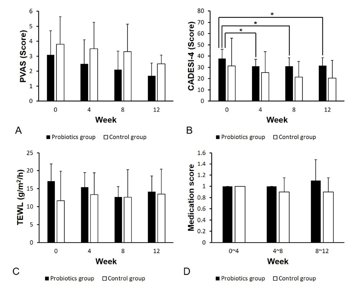
Although the PVAS scores in both groups consistently decreased until the end of the experiment, there were no significant within-group differences at all time-points (Fig. 1A). Further, there were no significant between-group differences at each time point (baseline, p = 0.694; 4 weeks, p = 0.280; 8 weeks, p = 0.336; 12 weeks, p = 0.197).
CADESI-4
Compared to the baseline scores, there was a significant decrease of the CADESI-4 scores at 4, 8, and 12 weeks in the probiotics group (0-4 weeks, p = 0.02; 0-8 weeks, p = 0.03; 0-12 weeks, p = 0.02) (Fig. 1B). However, there was no significant between-group difference at each time point (0 week, p = 0.824; 4 weeks, p = 0.715; 8 weeks, p = 0.291; 12 weeks, p = 0.382).
TEWL
There was no significant between-group difference at each time point (baseline, p = 0.158; 4 weeks, p = 0.158; 8 weeks, p = 0.842; 12 weeks, p > 0.999). In addition, there was no evident effect of the probiotics and placebo powder on TEWL within each group over time (Fig. 1C).
Medication score
Regarding the medication score, there was a slight increase at 8-12 weeks in the probiotics group and a slight decrease at 4-8 weeks in the control group; however, there were no significant within-group differences at all time-points (p > 0.99). Moreover, there were no significant between-group differences at each time point (p > 0.99).
Discussion
Several studies have reported beneficial effects of probiotics in the prevention and management of AD in human patients. Specifically, a study reported that infants of mothers who had received the probiotics had a reduced incidence rate of AD compared to the control group [17]. Further, administration of probiotics to children aged 6 months to 13 years and with moderate or severe AD has been reported to moderately improve the clinical severity of AD [8,9]. In adult AD patients, there have been reported improvements of cutaneous symptoms, intestinal flora in the feces, and patients' psychological status [10]. There have also been several studies using mouse model of AD. In these studies [18-20], probiotics prevented the development of AD and allergic march, progression of allergic disorders from AD, by inhibition of inflammatory cytokines.
In veterinary medicine, there have been few reports on the effects of probiotics on canine AD. Moreover, ICADA has suggested that there is currently insufficient evidence on the clinical basis for the administration of oral probiotics as nonspecific immunotherapy to treat or prevent canine AD [4]. However, in the last decade, several studies on the complementary effects of Lactobacilli on canine AD have been performed [5-7]. They indicated that probiotics showed complementary benefits for dogs with AD who had been managed with conventional treatment. Three of these studies were based on the experimental AD model sensitized by house dust mite and only one study on Lactobacillus paracasei K71 performed actual veterinary clinical trials. Although it is already known that the beneficial effects of probiotics are greatly dependent on the specific strains, e.g., Lactobacilli and Bifidobacteria [21], there has been no study on the effect of Bifidobacteria on canine AD.
Intestinal bacterial flora is important not only in retaining the structural and functional integrity of the intestine but also in immune system regulation [22,23]. There have been several attempts to use probiotics to control the canine intestinal bacterial flora since intestinal microbiota imbalance may affect the gut as well as extra-intestinal organ systems such as the skin [24,25]. The specific mechanisms through which the gut microbiota produces its effects are beginning to be understood with some studies reporting a close correlation between intestinal microflora and numerous factors associated with the pathophysiology of AD, such as immunity and inflammation [26,27].
Based on the results of the study using Lactobacilli strain [7], we examined the hypothesis that probiotics have a beneficial effect on the alleviation of canine AD using a different strain of probiotics, i.e., Bifidobacteria, instead of Lactobacilli. However, in this randomized, placebo-controlled study, we found clinical complementary effects of the addition of probiotics for controlling the severity of canine AD by alleviating skin lesions. We found that the CADESI-4 scores of the probiotics group significantly decreased from the baseline to 4, 8 and 12 weeks; however, this was not observed in the control group. These findings are consistent with those of a study evaluating the complementary effects of Lactobacillus paracasei K71 on canine AD [7]. TEWL in the probiotics group showed a similar tendency; however, it did not reach statistical significance within the group. Therefore, it appears that B. longum might have supplementary effect of only improving skin lesions in dogs with AD receiving standard therapy. However, this effect might be fairly restricted given that there was no statistically significant difference compared to the control group in the CADESI-4 scores after initiation of administration.
Although many human studies have reported a beneficial effect of probiotics in not only improving clinical signs of AD but also reducing the incidence of AD, other studies have reported that probiotics supplementation neither improved the severity of AD nor reduced the incidence of AD [28,29].
A possible association between probiotics use and some adverse effects in humans has been reported. Further, theoretical side effects such as systemic infections, deleterious metabolic activities, excessive immune stimulation in susceptible individuals, gene transfer, and minor gastrointestinal symptoms have been reported [30]. However, in this study, no adverse effects of probiotics were reported by the dogs-owners and veterinarians.
The limitation of this study is the relatively small size of dogs. Although we did not find a significant between-group difference in the clinical signs, studies with larger samples are needed to further evaluate the effect of probiotics. Moreover, since several studies have reported that administration of probiotics prevented the development of AD in mouse model [18-20], further research is needed to evaluate the effect of probiotics on pregnant and young adult dogs. Further, future studies should consider stopping the use of drugs for canine AD treatment so as to ideally identify the beneficial effects of probiotics. In this study, it was difficult for the dog-owners to visit the veterinary hospital without any medication for 12 weeks. Therefore, the dogs in this study were maintained with a minimum dose of medication to manage the clinical signs of AD.
In conclusion, although it was not sufficient to reduce pruritus, TEWL, and the dosage of prescribed drugs for managing canine AD, our preliminary results showed that oral administration of B. longum was effective in improving skin lesions and could be considered as a supportive therapy for canine AD.
 PDF Links
 PubReader
 ePub Link
 Full text via DOI
 Download Citation
 Print
Print


