Retrospective evaluation of toceranib phosphate (Palladia) for treatment of different tumor types in 31 dogs
Article information

Abstract
The purpose of this retrospective study was to provide additional data on the use of toceranib in a wide variety of tumor types in small breed dogs, especially < 8 kg (except 5 dogs). This was a retrospective study of 31 dogs with malignant tumors treated with a 2.5 mg/kg median dose of toceranib (Palladia; Zoetis, USA) on a Monday–Wednesday–Friday schedule. Clinical benefit was observed in 13 of 15 dogs (86.7%, 3 with complete response, 4 with partial response, 6 with stable disease) with gross disease. Distant metastasis, response to treatment, and treatment setting were significantly associated with survival time. Negative prognostic factors were multiple chemotherapy and distant metastasis (affecting progression-free survival [PFS]), surgery, regional enlarged lymph nodes, underlying disease, and toxicity (affecting median survival time [MST]). Positive prognostic factors were epithelial and round cell tumor (affecting PFS), epithelial tumor, microscopic disease, no evidence of disease response, and stable disease (MST). In conclusion, a clinical benefit from toceranib treatment was noted in most of the dogs with gross disease in our study. This study suggested that the toceranib is probably selective treatment to various tumor types in small breed dogs.
Introduction
Toceranib phosphate, a small molecule tyrosine kinase inhibitor, is commonly used in veterinary oncology. Receptor tyrosine kinases (RTKs) are good candidates for targeted molecular treatment as they play important roles in cell survival and proliferation and are dysregulated in a diverse range of malignancies through overexpression, activating mutations, and autocrine activation loops [1]. Toceranib inhibits both normal and mutated RTKs by competitive inhibition of adenosine triphosphate binding, which is need to phosphorylation and downstream signaling. The targets of toceranib are split-kinase family elements such as the FMS-like tyrosine kinase-3, KIT, vascular endothelial growth factor receptor 2, and platelet-derived growth factor receptor β, in a similar manner to sunitinib malate, another small-molecule inhibitor of RTKs [2,3]. Toceranib has not only antitumor but also antiangiogenic effects, and therefore has the potential to treat various tumor types [1,4-6].
Toceranib was approved by the Food and Drug Administration for the treatment of canine mast cell tumors (MCTs) in 2009. Its safety and activity were first assessed in a Phase 1 clinical trial involving a total of 57 dogs with a variety of tumors [5]. In this study, 31 dogs had an objective response (54.4%; 6 with complete response [CR], 10 with partial response [PR], and 15 with stable disease [SD]), demonstrating the potential biological activity of toceranib. The highest response rate was observed in 22 dogs with MCTs (59.1%, 13/22), including 11 dogs with KIT mutations (90.9%, 10/11) and sarcomas, carcinomas, melanomas, and myeloma also responded to the treatment [5]. Various reports have described the evaluation of toceranib [7,8]. Apart from MCTs [6,9], off-label uses have been described in a variety of tumors, including those of epithelial origin (apocrine gland anal sac adenocarcinoma [AGASA] [10,11], squamous cell carcinoma [SCC] [12], hepatocellular carcinoma [13], nasal carcinoma [11], thyroid carcinoma [14], and mammary gland tumor [15]), mesenchymal origin (osteosarcoma [OSA] [11,16,17], gastrointestinal stromal tumor [GIST] [18], and melanoma [5]), and round cell origin (lymphoma [19]). In one study that reported toceranib’s use in the treatment of solid tumors (AGASA, OSAs, thyroid carcinoma, and head and neck carcinoma), a clinical benefit was observed in 63/85 (74.1%) dogs [11].
Studies evaluated prognostic factors for the application of toceranib to general tumors were not enough and no cases were previously applied to majority of small breed dogs. Therefore, the purpose of the following retrospective study was to provide additional data on the use of toceranib in a wide variety of tumor types in small breed dogs, especially < 8 kg (except 5 dogs). This study describes the responses of toceranib treatment to various tumors, which has been reported for the first time in republic of Korea.
Materials and Methods
Case selection and treatment procedures
The client-owned dogs in this study were treated at the Veterinary Medical Teaching Hospital College of Chungnam National University or local referral veterinary clinics in Korea between January 2016 and September 2020. Medical charts with the owner’s consent to the use of patient information prior to medical care were used in this study. Authors declare no IACUC or other approval was needed for our study. We identified dogs that received diagnoses of malignant tumors by reviewing the medical records, from which we also recorded signalment (age, sex, and breed), physical examination, complete blood count (CBC), serum biochemistry profile, urinalysis, diagnostic imaging, histopathological assessment, toceranib dose and schedule (interval and duration), concomitant medications, and follow-up information including adverse events (AEs) and response to treatment. Lymph node (LN) assessment was performed in all patients through ultrasonography (US) and distant metastasis was evaluated by computed tomography (CT). In addition, the basal conditions of all patients were recorded before the treatment began. Dogs were excluded from the study (n = 7; 6 epithelial origin and 1 mesenchymal origin) if they were treated with toceranib for less than 28 days because it is difficult to assess the effects in short courses of treatment, as has been previously reported in solid tumors (except round cell populations) [1,16].
Re-evaluations, including physical examination, CBC, serum biochemistry, and urinalysis, were performed within a month of starting toceranib (usually within two weeks). Tumors were restaged based on direct measurement of gross mass or imaging (radiography, US, or CT) and this continued up to every 3 months periodically until toceranib ceased.
Toceranib (Palladia; Zoetis, Florham Park, NJ, USA) was administrated at a dose of 2.4 to 2.9 mg/kg which causes sufficient target inhibition and the treatment schedule was on a Monday–Wednesday–Friday basis in most dogs. In some cases, the dose and schedule were adjusted depending on patient’s condition [20]. In the case of symptoms of toxicity (lethargy, weight loss, inappetence, diarrhea, vomiting, fever, syncope), toceranib was discontinued until side effects improved, and then re-administration began with a dose reduced by 0.5 mg/kg.
Most of dogs (20/31) with surgical removal of tumor burden or biopsy were confirmed by histopathological examination. The surgical margin, mitotic count, and vascular invasion of tumor were evaluated for each malignant tumor according to the histopathology report. Some dogs (11/31) were tentatively diagnosed by cytology based on fine needle aspiration in mostly MCTs (6 of 11). Toceranib was administrated first by the choice of owner who was burdened with surgery or intravenous injection of antitumor drug or by the presence of distant metastasis or to prevent recurrence after surgery. Vinblastine and prednisolone with or without lomustine were used as the first dugs for MCT treatment and replaced or combined with toceranib due to side effects, no treatment response or recurrence of tumor. Also, electrochemotherapy was applied to gross tumor (relapsed or not) with toceranib. At the time of treatment, 10 dogs had other existing underlying diseases.
Response to therapy
The response to therapy was assessed in 31 dogs using diagnostic images (radiographs, US, or CT) and direct measurement of the tumor diameter if possible. The responses of gross (macroscopic) tumors in dogs without surgical intervention or with recurrence of resected tumor were defined as CR (complete regression of the target tumor, no new lesions), PR (partial regression of the target tumor, ≥30% decrease in the longest diameter of the target tumor, no new lesions), progressive disease (PD, > 20% increase in the longest diameter of the target tumor, progression of nontarget lesions and new lesions), or SD (SD, decrease of the target tumor of less than 30% or increase of the target tumors of less than 20%, no progression of nontarget lesions and no new lesions for at least 10 weeks) [21]. The responses of tumors in dogs with microscopic disease after surgical intervention were defined as no evidence of disease (NED, at initiation of treatment, no relapse) or PD (metastasis or recurrence of tumor that has been surgically removed or presence of new lesions).
Progression-free survival (PFS) was defined as the time from the initiation of toceranib to PD or death from any cause. The median survival time (MST) was defined as the time from initiation of toceranib to death from any cause. If the dog was alive at the time of writing this manuscript, the survival time was calculated using the last date recorded in the medical notes.
PFS and MST were assessed in the total population (31 dogs) by reviewing the medical records. Clinical benefit was determined by response to treatment and was defined as CR or PR of any duration, or SD for at least 10 weeks in dogs with gross disease.
Assessment of AEs
All dogs receiving toceranib were evaluated by physical examination, CBC, serum biochemistry, and urinalysis before treatment and then at intervals that gradually increased from 2 weeks to 1 to 3 months if AEs were not found. All AEs were classified by the attending clinician according to the Veterinary Co-operative Oncology Group’s common terminology criteria for AEs (VCOG-CTCAE v1.1) [22]. Multiple selection was allowed for each dog. For gastrointestinal AEs, supportive care included antiemetics, antidiarrheal agents, and gastric protectants. When neutropenia occurred, prophylactic antibiotics were prescribed until the next evaluation.
Statistical analysis
The Kaplan–Meier method was used to examine PFS and MST according to the studied characteristics of the dogs. Median PFS could not be estimated as tumors recurred in nine dogs (29.0%). Therefore, mean PFS was used. Cox regression analysis was performed to determine factors affecting the recurrence of tumor or death after initiation of toceranib treatment. Variables were selected by a backward elimination method. All statistical analyses were performed using IBM SPSS Statistics for Windows, version 25.0 (IBM Corp., USA). A p-value of < 0.05 was considered statistically significant.
Results
Study population
Thirty-one dogs with tumors were treated with toceranib either alone or in combination with metronomic chemotherapy, surgery, or both for the study period. The population included 21 female dogs (18 spayed) and 10 male dogs (8 castrated). Fourteen different breeds were presented (Table 1). The median age was 13 years (range, 6 to 18 years) and the median body weight was 5 kg (range 2.3 to 42 kg). The types of tumor characterized in the study population consisted of epithelial tumors (n = 12), mesenchymal tumors (n = 10), and round cell tumors (n = 9). The majority of the dogs (51.6%) were treated with toceranib in the absence of evidence of mass after surgical resection of a tumor. The remaining dogs (48.4%) were treated in the presence of primary tumor, metastasis, recurrent tumor, or a combination thereof. Metastasis located in distant organs was present in only 3 dogs (9.7%) at initiation of toceranib, but 14 dogs (45.2%) had enlarged LNs around the target tumor. Detailed patient characteristics are presented in Table 2.
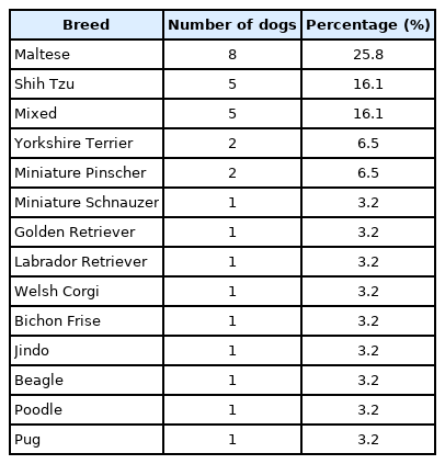
Summary of breeds included in this study (n = 31)
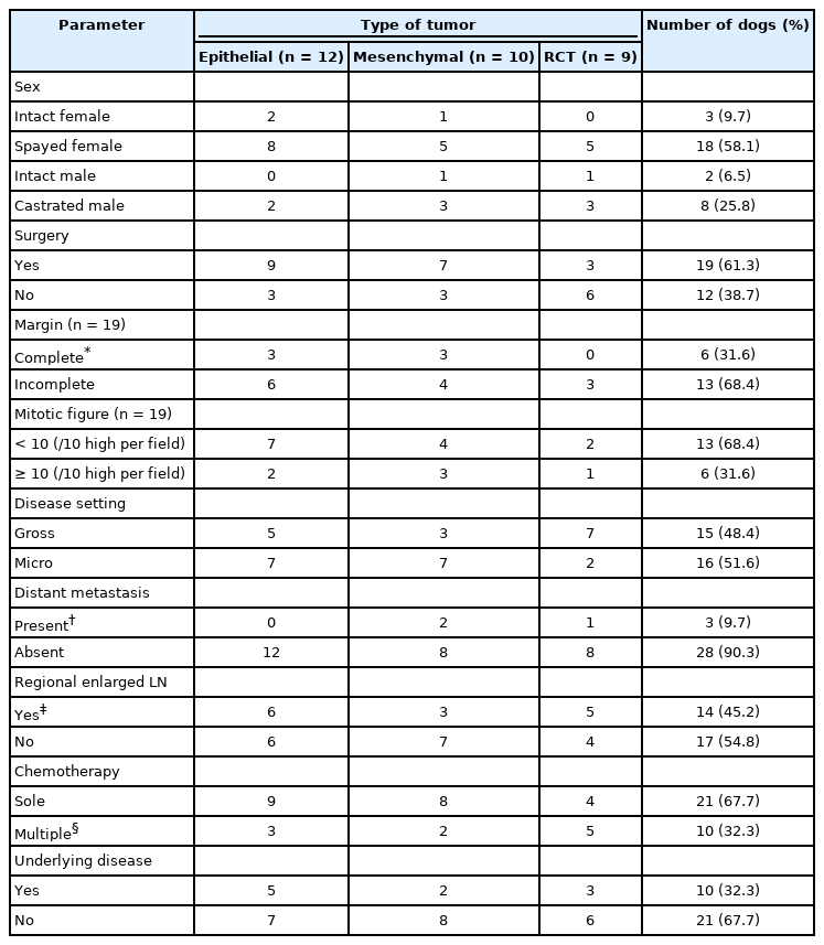
Patient characteristics and additional tumor information (n = 31)
Overview table with following information for each dog enrolled in the study was described in the supplement.
Treatments
The median dose of toceranib used in treated dogs was 2.5 mg/kg (range, 2.2 to 3 mg/kg). Most dogs (87.1%, 27/31) were administered toceranib three days per week (Monday–Wednesday–Friday), but one dog was administered toceranib every second day and three dogs two days per week (Monday–Thursday).
Nineteen dogs (61.3%) underwent surgery before initiation of treatment. In four of these dogs, regional enlarged LNs removed during surgery were confirmed to have metastases by histopathology. An additional ten dogs had regional enlarged LNs suspected to have metastases after CT or US. Three dogs (9.7%) had distant metastasis of lungs or abdominal carcinomatosis. Histopathologic details such as margin and mitotic count were well characterized (Table 3). There was no vascular invasion of tumor in any of the dogs examined.

Clinical benefits of toceranib administration in 15 dogs with gross disease
Of the 31 dogs in the study population, 21 were treated solely with toceranib and 10 were treated with other chemotherapies during toceranib treatment. Other chemotherapy agents included electrochemotherapy with bleomycin (0.3 mg/kg), intratumor injection (n = 3; SCC, intraperitoneal liposarcoma, MCT), vinblastine (n = 3; MCTs), carboplatin (n = 1; SCC), prednisolone (n = 5; four MCT, one MGT), and non-steroidal anti-inflammatory drugs (n = 2; SCC, MGT).
Treatment outcome
Clinical benefit was observed in 13 of 15 dogs (86.7%; 3 with CR, 4 with PR, 6 with SD) with gross disease. The remaining two dogs (13.3%) had PD (Table 3). Of the 13 dogs for which toceranib treatment showed benefit, 11 presented prolonged clinical benefit (median 35 days; range, 7 to 490 days) from cessation of toceranib to death or last date reported. 2 dogs in these groups were expired during toceranib treatment; 1 had PR, which then turned to PD, and 1 with SD died due to deterioration from toceranib-related side effects. The 16 dogs (51.6%) with microscopic disease were defined as having NED at the initiation of toceranib treatment. Seven dogs with NED had PD during toceranib treatment or after discontinuation of toceranib.
In the 31 treated dogs, the toceranib-related mean PFS and median MST were 471 and 199 days, respectively, based on the Kaplan–Meier product of survival time (Fig. 1). Distant metastasis was significantly associated with PFS (p < 0.05). Response to treatment and treatment setting (macro- or micro-scopic) were significantly associated with MST (p < 0.001 and p < 0.05, respectively). The median MSTs for dogs with CR, PR, SD, and PD were 311, 133, 187, and 156 days, respectively. The mean PFS and median MST were 440 and 290 days, respectively, for dogs that underwent surgery and 450 and 154 days, respectively, for dogs that did not undergo surgery. The mean PFS and median MST were 315 and 199 days, respectively, for dogs with mesenchymal tumors, 588 and 311 days, respectively, for dogs with epithelial tumors, and 421 and 156 days, respectively, for dogs with round cell tumors. The mean PFS and median MST were 410 and 290 days, respectively, for dogs with microscopic disease, compared with 463 and 169 days, respectively, for dogs with gross disease. The mean PFS and median MST were 389 and 169 days, respectively, for dogs that received multiple chemotherapeutic drugs, compared with 463 and 289 days, respectively, for dogs that received toceranib alone. The mean PFS and median MST were 90 and 187 days, respectively, for dogs with distant metastasis, compared with 493 and 220 days, respectively, for dogs with no distant metastasis. The mean PFS and median MST were 224 and 169 days, respectively, for dogs with regional enlarged LNs, compared with 553 and 289 days, respectively, for dogs which did not have regional enlarged LNs. The mean PFS and median MST were 425 and 187 days, respectively, for dogs with underlying disease, compared with 413 and 289 days, respectively, for dogs without underlying disease. The mean PFS and median MST were 469 and 289 days, respectively, for dogs with symptoms of toxicity to toceranib, compared with 388 and 199 days, respectively, for dogs without toxicity to toceranib (Table 4).
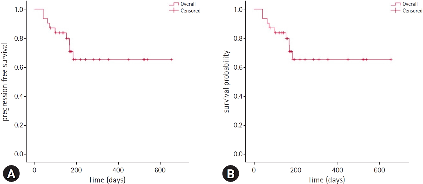
(A) Kaplan–Meier progression free survival curve based on the time of toceranib initiation (mean 471 days, range 370–571); (B) Kaplan–Meier median survival time curve based on the time of toceranib treatment initiation (median 199 days, range 89–309). All censored dogs are marked with a cross.

Progression-free survival (PFS) and median survival time (MST) based on the Kaplan–Meier method
Prognostic factors
Multiple factors were assessed for their influence on prognosis using a multivariable Cox proportional hazard model for PFS (Table 5) and MST (Table 6). Multiple chemotherapy and distant metastasis (in PFS) and surgery, regional enlarged LN, underlying disease, and toxicity (in MST) negatively affected prognosis. Conversely, epithelial and round cell tumors and duration of toceranib (in PFS) and epithelial tumors, microscopic disease, NED, SD, and duration of toceranib (in MST) positively affected prognosis.
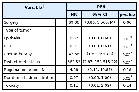
Prognostic factors based on Cox proportional hazard model for progression-free survival (PFS)

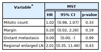
Prognostic factors based on Cox proportional hazard model for median survival time (MST)
In dogs that underwent surgery, none of the examined factors were significantly associated with MST (Table 7).
Prognostic factors based on Cox proportional hazard model for median survival time (MST) in the group that underwent surgery
Adverse events
AEs occurred in 14/31 (45.2%) of the dogs treated with toceranib. The categories, grades (1 to 3), and incidence rate per category of these AEs are described in Table 8. There were no grade 4 or 5 AEs observed in any dogs. The majority of the AEs was grade 1 (54.0%) and categorized as biochemical (39.7%) or gastrointestinal (28.6%) AEs. Most of the gastrointestinal AEs (8/14) were classified as low grade (grades 1 to 2) and were managed quickly by supportive therapy with discontinuation of toceranib. In 6/14 (42.9%) dogs, toceranib administration was discontinued due to AEs (3 biochemical: increased blood urea nitrogen and creatinine; 2 hematologic: 1 neutropenia and 1 anemia; 1 gastrointestinal).
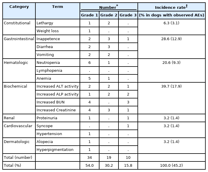
Adverse events (AEs) in dogs treated with toceranib (45.2% of the total study cohort)
Discussion
This retrospective study describes the responses of toceranib treatment to various tumors, which has been reported for the first time in republic of Korea. The biological effect of toceranib on various types of tumors was evaluated in mostly small breed dogs. Most of the treated dogs with gross disease (48.4% of the total cohort) presented with clinical benefit (86.7%). This treatment response is consistent with previous studies [10,11,18]. Most previous reports tested the effect of toceranib on one type of tumor while our study supports the use of toceranib in various types of tumor. There have been few studies of the relationship between tumor type and prognosis, but our study shows that epithelial tumors have a low recurrence rate and a high survival rate. In the previous studies assessing toceranib, clinical benefits have mainly been shown in epithelial tumors (‘-carcinoma’) and MCTs [1,6,10,11,13,14,18]. Conversely, few studies have reported effects on mesenchymal tumors such as GIST, OSA, injection site sarcoma, and histiocytic sarcoma, and these studies were limited by small populations [11,18,23-25]. Mutations in c-KIT, an important factor which is the target of small-molecule tyrosine kinase inhibitors in veterinary medicine, have been found in MCTs (grades 1 to 3) in dogs, and in GIST and mammary gland tumor (grades 2 to 3) in dogs and humans [15,26-28]. In our study, dogs with epithelial tumors with sufficient clinical evidence had the highest representation (12 of 31 dogs, 38.7%) and showed clinical benefit from toceranib treatment, including two dogs with CR.
Using a Cox regression analysis, we identified that NED or SD response to treatment had a significantly positive effect on prognosis (in MST). Unexpectedly, CR and PR did not have a significant effect on prognosis, despite the clinical benefit of toceranib. A previous report showed that PR and SD were positive factors of prognosis [9]. Since our study involved 31 dogs and was limited to three centers, a direct comparison with previous retrospective studies may not be appropriate. However, the following points may explain why the effect of PR on prognosis found in our study differed from that in other studies. In our study, a total of four dogs (one with SCC, and three with MCT, stage 3) that experienced PR all died of each tumor, and their median MST was 133 days. Of these, the first responded to secondary toceranib treatment after several trials with other chemotherapies, but had recurrence two weeks before death and the patient’s condition was deteriorated quickly. The second dog also experienced recurrence two weeks before death from a distant metastasis (carcinomatosis). The third dog initially showed PR, but the tumor continued to increase, and leading to PD. The fourth was already in a state of relapse after surgery when toceranib was initiated. Therefore, the dogs that experienced PR all showed rapid relapse and deterioration of patient’s condition even though a clinical benefit from the treatment was recorded. Overall, the fact that our study had conditions differing from the earlier report needs to be fully considered and interpreted.
In our study, dogs that underwent surgery had a worse prognosis than those that did not, and dogs with microscopic disease had a better prognosis than those without. In a previous report, the effect of surgery on prognosis was not statistically significant [10]. In our study, of the dogs that underwent surgery, 16/19 initiated toceranib treatment in a microscopic disease setting and recurrence occurred in three dogs after surgery, resulting in gross disease (3/19). Considering the result that the microscopic condition had a significant positive effect on the prognosis, these dogs relapsed after surgery had poor MST, even if clinical benefit (two with CR and one with PR) was observed. So, surgery was considered a negative factor of prognosis in our study.
In dogs with regional enlarged LN and distant metastasis, the prognosis (MST) was poor. Previous studies have shown results consistent with this finding [10,17,18,29]. Conversely, in one report related to grade 2 MCTs, the survival time between dogs with and without regional LN metastasis was no significant difference. However, prolonged survival time was observed in dogs in which metastatic LNs were removed [30]. Our study identified regional enlarged LN was removed in only 4 of 14 dogs.
The median dose of toceranib used in treated dogs was 2.5 mg/kg (range, 2.2 to 3 mg/kg). According to existing reports, low doses of 2.5 to 2.9 mg/kg every other day are well tolerated without grade 3 or 4 AEs, compared with dosing at 3.25 mg/kg every other day [20]. In our study, 10 dogs had existing underlying diseases (three with heart failure, one with chronic kidney disease, four with both heart and kidney disease, and two with chronic pancreatitis). Depending on these concomitant diseases, main side effects were occurred and therefore biochemistry AEs was the most common (39.7%), unlikely to another reports. Gastrointestinal AEs were the second most common type of AEs, and these were effectively managed in grades 1 to 2 through drug holidays, supportive medicine, and dose adjustments to avoid drug withdrawal. The dogs that died from severe side effects developed worsening chronic kidney disease, severe pancreatitis, or anemia and did not recover.
There were several limitations of our study. Not only was the overall population size small, but the numbers in the subgroup analyses were also small. These findings should be further confirmed in a large number of small breed dogs. As this study had a retrospective design, it was impossible to match baseline characteristics such as sex, biochemistry, and underlying diseases. Additionally, toceranib affecting the tumor microenvironment (TME) in various tumor types may be assessed by molecular biology in other ways, such as Western blots or immunohistochemistry [31].
In conclusion, a clinical benefit of toceranib treatment was noted in the majority of the 15 dogs with gross disease in our study (86.7%). In the 31 treated dogs, the toceranib-related mean PFS was 471 days and the median MST was 199 days. Complex chemotherapy and distant metastasis (PFS), surgery, regional enlarged LN, underlying disease, and toxicity (MST) were negative prognostic factors. Our results provide additional information about the clinical efficacy of toceranib and suggest that the toceranib is probably selective treatment to various tumor types in small breed dogs. Further study is required to evaluate its clinical activity in a large number of small breed dogs.
Notes
The authors declare no conflict of interest.
Acknowledgements
We are thanks to the Cooperative Research Program of Center for Companion Animal Research (Project no. PJ014045022020): Rural Development Administration, Republic of Korea for the support.
Supplementary Materials
Overview table with additional information for each dog enrolled in the study (n = 31).
Overview table with additional information for each dog enrolled in the study (n = 31).