


ņä£ļĪĀ
Fenbendazole (FBZ)ņØĆ ņłśņØśņŚÉņä£ ņ£ĀņÜ®ĒĢśĻ▓ī ņō░ņØ┤ļŖö ĻĄ¼ņČ®ņĀ£ ņżæ ĒĢśļéśņØ┤ļŗż[1]. FBZņØĆ ņŗżĒŚśņÜ® ņäżņ╣śļźś, ļ░śļĀżļÅÖļ¼╝, ņé░ņŚģļÅÖļ¼╝ņŚÉņä£ ĒÜīņČ®, ņŗŁņØ┤ņ¦ĆņןņČ®, ĒÄĖņČ® ļō▒ ļé┤ļČĆ ĻĖ░ņāØņČ® Ļ░ÉņŚ╝ņØś ņśłļ░® ļ░Å ņ╣śļŻīņĀ£ļĪ£ ĒŚłĻ░Ć ļ░øņĢä ņé¼ņÜ®ļÉśĻ│Ā ņ׳ļŗż[2,3]. ĻĄ¼ņČ® ņ×æņÜ®ĻĖ░ņĀäņ£╝ļĪ£ļŖö ĻĖ░ņāØņČ®ņØś ņäĖĒż ļČäņŚ┤ Ļ│╝ņĀĢ ņżæ ļ»ĖņäĖņåīĻ┤ĆņØś ĒśĢņä▒ņØä ļ░®ĒĢ┤ĒĢśņŚ¼ ĒÜ©Ļ│╝ļź╝ ļéśĒāĆļé┤ļŖö Ļ▓āņ£╝ļĪ£ ņĢīļĀżņĀĖ ņ׳ļŗż[4,5].
ņĄ£ĻĘ╝ FBZņØ┤ ĒĢŁņĢöĒÜ©Ļ│╝Ļ░Ć ņ׳ļŗżļŖö ļ│┤Ļ│Ā[6]ņÖĆ ņØ╝ļČĆ ņ×äņāüņé¼ļĪĆļź╝ ņŗ£ņ×æņ£╝ļĪ£ ņ╗żļ«żļŗłĒŗ░, ņØĖĒä░ļäĘņØä ĒåĄĒĢ┤ FBZņØä ņ×äņØśļĪ£ ļ│ĄņÜ®ĒĢśļŖö ņé¼ļĪĆĻ░Ć ņ”ØĻ░ĆĒĢśņśĆļŗż. FBZņØĆ ņØĖĻ░ä ņĢöņäĖĒżņØś ļ»ĖņäĖņåīĻ┤ĆņŚÉ ņ╣£ĒÖöņä▒ņØä ļéśĒāĆļé┤Ļ│Ā micromolar ļåŹļÅäņŚÉņä£ ĒĢŁņĢöĒÜ©Ļ│╝ļź╝ ļ│┤ņśĆņ£╝ļ®░, ņāłļĪ£ņÜ┤ ĒĢŁņĢöĻĖ░ņĀäņ£╝ļĪ£ GLUT transporterņÖĆ hexokinaseņØś ļ░£Ēśä ņ¢ĄņĀ£ļĪ£ glucose uptakeļź╝ ĒÜ©Ļ│╝ņĀüņ£╝ļĪ£ ņ¢ĄņĀ£ĒĢśļŖö ņé¼ņŗżņØ┤ ņĢīļĀżņĪīļŗż[6].
Ļ│©ņłśļŖö ĒśłņĢĪņäĖĒżņÖĆ ļ®┤ņŚŁņäĖĒżĻ░Ć ņ£ĀļלĒĢśļŖö ĻĖ░Ļ┤Ćņ£╝ļĪ£ ļ®┤ņŚŁņ▓┤Ļ│äņŚÉņä£ ļ¦żņÜ░ ņżæņÜöĒĢ£ ņŚŁĒĢĀņØä ĒĢ£ļŗż. ņÜ░ļ”¼ļŖö FBZņØ┤ ĒĢŁņĢöņĀ£ļĪ£ņä£ņØś Ļ░ĆļŖźņä▒ņØ┤ ļīĆļæÉļÉśĻ│Ā ņ׳ļŖö Ļ░ĆņÜ┤ļŹ░ Ļ│©ņłśņäĖĒżņŚÉ ļīĆĒĢ£ ĒĢŁņŚ╝ņ”Ø ĒÜ©Ļ│╝ ļśÉĒĢ£ ņ׳ļŖöņ¦Ć ņØśļ¼ĖņØ┤ ļōżņŚłļŗż. ĒĢśņ¦Ćļ¦ī ņĢ×ņäĀ ņŚ░ĻĄ¼ņŚÉņä£ļŖö ņĪ░ļźśņŚÉņä£ ņäĀņČ® Ļ░ÉņŚ╝ņ£╝ļĪ£ ņØĖĒĢ┤ ļ░£ņāØĒĢ£ Ļ│©ņłśņŚ╝ņŚÉ FBZņØä Ēł¼ņŚ¼ĒĢśņŚ¼ ņ╣śļŻīĒĢ£ ņé¼ļĪĆļ¦īņØ┤ ņĪ┤ņ×¼ĒĢśņśĆļŗż[7]. ņØ┤ņÖĆ Ļ░ÖņØ┤, FBZņØś Ļ│©ņłśņŚ╝ņŚÉ ļīĆĒĢ£ ĒÜ©Ļ│╝ņŚÉ Ļ┤ĆĒĢ£ ņŚ░ĻĄ¼ļŖö ļ¦ÄņØ┤ ļČĆņĪ▒ĒĢśļŗż. ļö░ļØ╝ņä£ ļ│Ė ņŚ░ĻĄ¼ņŚÉņä£ļŖö in vitroņŚÉņä£ Ļ│©ņłśņŚ╝Ļ│╝ ņ£Āņé¼ĒĢ£ ņāüĒÖ®ņØä ļ¦īļōżĻĖ░ ņ£äĒĢ┤ ļīĆĒæ£ņĀüņØĖ ņŚ╝ņ”Ø ņ£Āļ░£ļ¼╝ņ¦łņØĖ lipopolysaccharide (LPS)ļź╝ Ļ│©ņłśņäĖĒżņŚÉ ņ▓śļ”¼ĒĢśņŚ¼ FBZņØś ĒĢŁņŚ╝ņ”Ø ĒÜ©Ļ│╝ ņ£Āļ¼┤ņÖĆ ņ×æņÜ®ĻĖ░ņĀäņØä ņĢīņĢäļ│┤ņĢśļŗż. ņØ┤ļź╝ ņ£äĒĢ┤ FBZņØä ļåŹļÅäļ│äļĪ£ ņ▓śļ”¼ĒĢ£ Ēøä Ļ│©ņłśņäĖĒżņØś ļīĆņé¼ĒÖ£ņä▒ļÅä, Ēæ£ļ®┤ ļ¦łņ╗ż ļ░£Ēśä, ņäĖĒżņØś ĒĢĄ ĒśĢĒā£, ļ»ĖĒåĀņĮśļō£ļ”¼ņĢä ļ¦ēņĀäņ£ä(mitochondrial membrane potential) ļō▒ņØä ņĖĪņĀĢĒĢśņŚ¼ Ļ│©ņłśņäĖĒżņŚÉņä£ FBZņØś ĒĢŁņŚ╝ņ”Ø ĒÜ©Ļ│╝ņŚÉ ļīĆĒĢ┤ ņŚ░ĻĄ¼ĒĢśņśĆļŗż.
ņ×¼ļŻī ļ░Å ļ░®ļ▓Ģ
ņŗżĒŚśļÅÖļ¼╝Ļ│╝ ņŗ£ņĢĮ
ņŗżĒŚśļÅÖļ¼╝ņØĆ OrientBio (Korea)ņŚÉņä£ ĻĄ¼ņ×ģĒĢśņŚ¼ ņĀ£ņŻ╝ļīĆĒĢÖĻĄÉ ņŗżĒŚśļÅÖļ¼╝ņä╝Ēä░ņŚÉņä£ ņ£Āņ¦ĆĒĢśņśĆļŗż. ļÅÖļ¼╝ņŗżĒŚśņŚÉņä£ 8-12ņŻ╝ļĀ╣ ņé¼ņØ┤ņØś C57BL/6 ļ¦łņÜ░ņŖżĻ░Ć ņé¼ņÜ®ļÉśņŚłĻ│Ā, ņĀ£ņŻ╝ļīĆĒĢÖĻĄÉ ļÅÖļ¼╝ņŗżĒŚśņ£żļ”¼ņ£äņøÉĒÜīņØś ņŖ╣ņØĖņØä ļ░øņĢä ņŗ£Ē¢ēļÉśņŚłļŗż(ņŖ╣ņØĖļ▓łĒśĖ, 2018-0011). FBZĻ│╝ LPS (Escherichia coli O55)ļŖö Sigmaņé¼(USA)ņŚÉņä£ ĻĄ¼ņ×ģĒĢśņśĆņ£╝ļ®░, FBZņØĆ dimethyl sulfoxide, LPSļŖö ņØĖņé░ņÖäņČ®ņĢĪņŚÉ ļģ╣ņØĖ Ēøä ņé¼ņÜ®ĒĢśņśĆļŗż.
Ļ│©ņłśņäĖĒżņØś ļČäļ”¼ņÖĆ ļ¼╝ņ¦ł ņ▓śļ”¼
ļ│Ė ņŗżĒŚśņŗżņŚÉņä£ ĒÖĢļ”ĮļÉ£ ļ░®ļ▓Ģņ£╝ļĪ£ Ļ│©ņłśņäĖĒżļź╝ ļČäļ”¼ĒĢśņśĆļŗż[8]. CO2 gasļĪ£ ļ¦łņÜ░ņŖżļź╝ ņĢłļØĮņé¼ ņŗ£Ēé© ļÆż ļīĆĒć┤Ļ│©Ļ│╝ Ļ▓ĮĻ│©ņØä ņĀüņČ£ĒĢśņŚ¼ Ļ│©ņłśņĪ░ņ¦üņØä ņ▒äņĘ©ĒĢśņśĆļŗż. ņØ┤ ņĪ░ņ¦üņØä ammonium chloride-potassium lysis bufferļĪ£ ņ▓śļ”¼ĒĢśņŚ¼ ņĀüĒśłĻĄ¼ļź╝ ņĀ£Ļ▒░ĒĢ£ Ēøä 70 ┬Ąm cell strainerņŚÉ Ļ▒Ėļ¤¼ single cellņØä ĒÜŹļōØĒĢśņśĆļŗż. ņØ┤ļź╝ Ļ│äņłśĒĢśņŚ¼ 96- ļśÉļŖö 6-well culture platesņŚÉ ļ░░ņ¢æĒĢśņśĆļŗż. FBZĻ│╝ LPSļź╝ ņ▓śļ”¼ĒĢ£ Ēøä 37Ōäā, 5% CO2ņØś ņĪ░Ļ▒┤ņŚÉņä£ ļ░░ņ¢æĒĢśņśĆĻ│Ā ļČäņäØņŚÉ ņØ┤ņÜ®ĒĢśņśĆļŗż.
Ļ│©ņłśņäĖĒżņØś ļīĆņé¼ĒÖ£ņä▒ļÅä ņĖĪņĀĢ
Ļ│©ņłśņäĖĒżļź╝ 1 ├Ś 106 cells/mLņØś ļåŹļÅäļĪ£ 96-well culture plateņŚÉ ļäŻņØĆ Ēøä FBZĻ│╝ LPS (1 ┬Ąg/mL)ļź╝ ļåŹļÅäļ│äļĪ£ ņ▓śļ”¼ĒĢ£ Ēøä ļ░░ņ¢æĒĢśņśĆļŗż. ļ░░ņ¢æņØ┤ ļüØļé£ Ēøä Ļ│©ņłśņäĖĒżņŚÉ 3-(4,5-dimethylthiazol-2-yl)-2,5-diphenyltetrazoliumbromide (MTT, Sigma) ņÜ®ņĢĪņØä 0.5 mg/mL ļåŹļÅäļĪ£ ļäŻĻ│Ā 4ņŗ£Ļ░ä ļÅÖņĢł ņ▓śļ”¼ĒĢśņśĆļŗż[9]. ņé┤ņĢäņ׳ļŖö ņäĖĒżņŚÉ ņØśĒĢ┤ ņāØĻĖ┤ crystal violetņØä ļģ╣ņØ┤ĻĖ░ ņ£äĒĢ┤ 10% sodium dodecyl sulfate ņÜ®ņĢĪņØä wellļŗ╣ 100 ╬╝Lņö® ļäŻņ¢┤ 2ņŗ£Ļ░ä ļÅÖņĢł ļ░śņØæņŗ£ņ╝░ļŗż. ĻĘĖ Ēøä microplate reader (Molecular Devicesņé¼, USA)ļź╝ ņØ┤ņÜ®ĒĢśņŚ¼ ĒØĪĻ┤æļÅä(570 nm)ļź╝ ņĖĪņĀĢĒĢśņśĆļŗż.
ņ£ĀņäĖĒż ļČäņäØ
ļ¦łņÜ░ņŖż Ļ│©ņłśņäĖĒżļź╝ 6-well culture plateņŚÉ 1 ├Ś 106 cells/mLņØś ļåŹļÅäļĪ£ ļ░░ņ¢æĒĢśĻ│Ā FBZĻ│╝ LPSļź╝ ļåŹļÅäļ│äļĪ£ ņ▓śļ”¼ĒĢśņśĆļŗż. 3ņØ╝Ļ░ä ļ░░ņ¢æ Ēøä ļ»ĖĒåĀņĮśļō£ļ”¼ņĢä ļ¦ēņĀäņ£äļź╝ ņĖĪņĀĢĒĢśĻĖ░ ņ£äĒĢ┤ rhodamine 123 ņÜ®ņĢĪņØä 10 ╬╝g/mLņØś ļåŹļÅäļĪ£ ņĢöņŗżņĪ░Ļ▒┤ņŚÉņä£ 30ļČäĻ░ä ņŚ╝ņāēĒĢśņśĆļŗż. Ļ│©ņłśņäĖĒżņØś ņäĖĒżņé¼(cell death) ņĖĪņĀĢņØä ņ£äĒĢ┤ annexin Ōģż-fluorescein isothiocyanate (FITC)ņÖĆ propidium iodide (PI) ņÜ®ņĢĪņ£╝ļĪ£ ņŚ╝ņāēĒĢśņśĆļŗż[10]. ļśÉĒĢ£, Ļ│╝ļ”ĮĻĄ¼ņÖĆ Bļ”╝ĒöäĻĄ¼ņØś ļ╣äņ£©ņØä ņĖĪņĀĢĒĢśĻĖ░ ņ£äĒĢ┤ allophycocyanin-labeled anti-Gr-1 ĒĢŁņ▓┤ņÖĆ biotin-labeled anti-B220 ĒĢŁņ▓┤, FITC-avidinņØä ņé¼ņÜ®ĒĢśņśĆļŗż. ņ£ĀņäĖĒż ļČäņäØņØĆ CytoFLEXņÖĆ CytExpert software (Beckman Coulter, USA)ļź╝ ņØ┤ņÜ®ĒĢ┤ ļČäņäØĒĢśņśĆļŗż.
Hoechst 33342 ņŚ╝ņāēņØä ņØ┤ņÜ®ĒĢ£ Ļ│©ņłśņäĖĒż ĒĢĄ Ļ┤Ćņ░░
Ļ│©ņłśņäĖĒżņØś ņäĖĒżņé¼ļź╝ ĒÖĢņØĖĒĢśĻĖ░ ņ£äĒĢ┤ ĒĢĄ ļ¬©ņ¢æņØä Ļ┤Ćņ░░ĒĢśņśĆļŗż. ņäĖĒżņØś ĒĢĄņØä ņŚ╝ņāēĒĢśĻĖ░ ņ£äĒĢ┤ Hoechst 33342 ņÜ®ņĢĪņØä 2.5 ╬╝g/mL ļåŹļÅäļĪ£ ņ▓śļ”¼ĒĢśņŚ¼ 37┬░CņŚÉņä£ 10ļČäĻ░ä ņŚ╝ņāēĒĢśņśĆļŗż[11]. ņŚ╝ņāēļÉ£ ņäĖĒżļŖö ĒśĢĻ┤æĒśäļ»ĖĻ▓Į(ZOE Fluorescent Cell Imager; BIO-RAD, USA)ņØä ņØ┤ņÜ®ĒĢ┤ Ļ┤Ćņ░░ĒĢśņśĆĻ│Ā ņé¼ņ¦ä ņ┤¼ņśüņØä Ē¢łļŗż.
ĒåĄĻ│äļČäņäØ
Ļ░ü ņŗżĒŚśņØĆ 2-3ĒÜī ļ░śļ│ĄĒĢśņŚ¼ ņŗ£Ē¢ēļÉśņŚłļŗż. Fig. 1ņØĆ ĒÅēĻĘĀ ┬▒ Ēæ£ņżĆĒÄĖņ░©ļĪ£ ļéśĒāĆļāłļŗż. One way analysis of variance ļČäņäØ ĒøäņŚÉ Turkey-Kramer multiple comparison test (GraphPad Prism; GraphPad Software, USA)ļĪ£ ņ£ĀņØśņä▒ņØä ĒÖĢņØĖĒĢśņśĆļŗż. ĒåĄĻ│äņ▓śļ”¼ Ēøä p-valueĻ░Ć 0.05 ļ»Ėļ¦īņØĖ Ļ▓ĮņÜ░ ņ£ĀņØśĒĢ£ Ļ▓āņ£╝ļĪ£ ĒīÉļŗ©ĒĢśņśĆļŗż.
Ļ▓░Ļ│╝
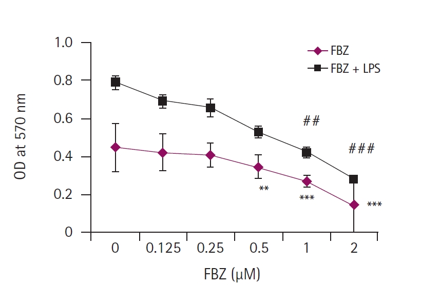
FBZņØ┤ Ļ│©ņłśņäĖĒżņØś ļīĆņé¼ĒÖ£ņä▒ļÅäņŚÉ ļ»Ėņ╣śļŖö ņśüĒ¢ź
FBZ ļŗ©ļÅģĻ│╝ FBZ + LPS (1 ╬╝g/mL) ņ▓śļ”¼ĻĄ░ņŚÉņä£ Ļ│©ņłśņäĖĒżņØś ļīĆņé¼ĒÖ£ņä▒ļÅäļź╝ ņĢīņĢäļ│┤ĻĖ░ ņ£äĒĢ┤ MTT assayļź╝ ņłśĒ¢ēĒĢśņśĆļŗż(Fig. 1). MTT assay Ļ▓░Ļ│╝ FBZ ļŗ©ļÅģņŚÉ ļ╣äĒĢ┤ FBZ + LPS ņ▓śļ”¼ĻĄ░ņØĆ ļ¬©ļōĀ ļåŹļÅä(0-1 ┬ĄM)ņŚÉņä£ ļ│┤ļŗż ļåÆņØĆ ļīĆņé¼ĒÖ£ņä▒ļÅäļź╝ ļéśĒāĆļé┤ņŚłņ£╝ļ®░, FBZņØś ļåŹļÅäĻ░Ć ņ”ØĻ░ĆĒĢ©ņŚÉ ļö░ļØ╝ ļīĆņé¼ĒÖ£ņä▒ļÅäĻ░Ć Ļ░ÉņåīļÉ©ņØä ĒÖĢņØĖĒĢśņśĆļŗż. ĒŖ╣Ē׳, FBZ ļŗ©ļÅģņØś 0.5-2 ┬ĄM ļåŹļÅäņŚÉņä£ ņ£ĀņØśĒĢśĻ▓ī ļīĆņé¼ĒÖ£ņä▒ļÅäĻ░Ć Ļ░ÉņåīĒĢśņśĆĻ│Ā, FBZ + LPS ņ▓śļ”¼ĻĄ░ņŚÉņä£ļŖö 1-2 ┬ĄM ļåŹļÅäņŚÉņä£ ņ£ĀņØśĒĢ£ Ļ░Éņåīļź╝ ļ│┤ņśĆļŗż. ņØ┤ļ¤¼ĒĢ£ Ļ▓░Ļ│╝ļŖö FBZņØ┤ LPSņÖĆ Ļ░ÖņØĆ ņŚ╝ņ”Ø ļ¼╝ņ¦łņŚÉ ņØśĒĢ┤ ņ×ÉĻĘ╣ļÉ£ Ļ│©ņłśņäĖĒżņØś ĒÖ£ļÅÖņØä ņ¢ĄņĀ£ĒĢĀ ņłś ņ׳ņØīņØä ļéśĒāĆļéĖļŗż.
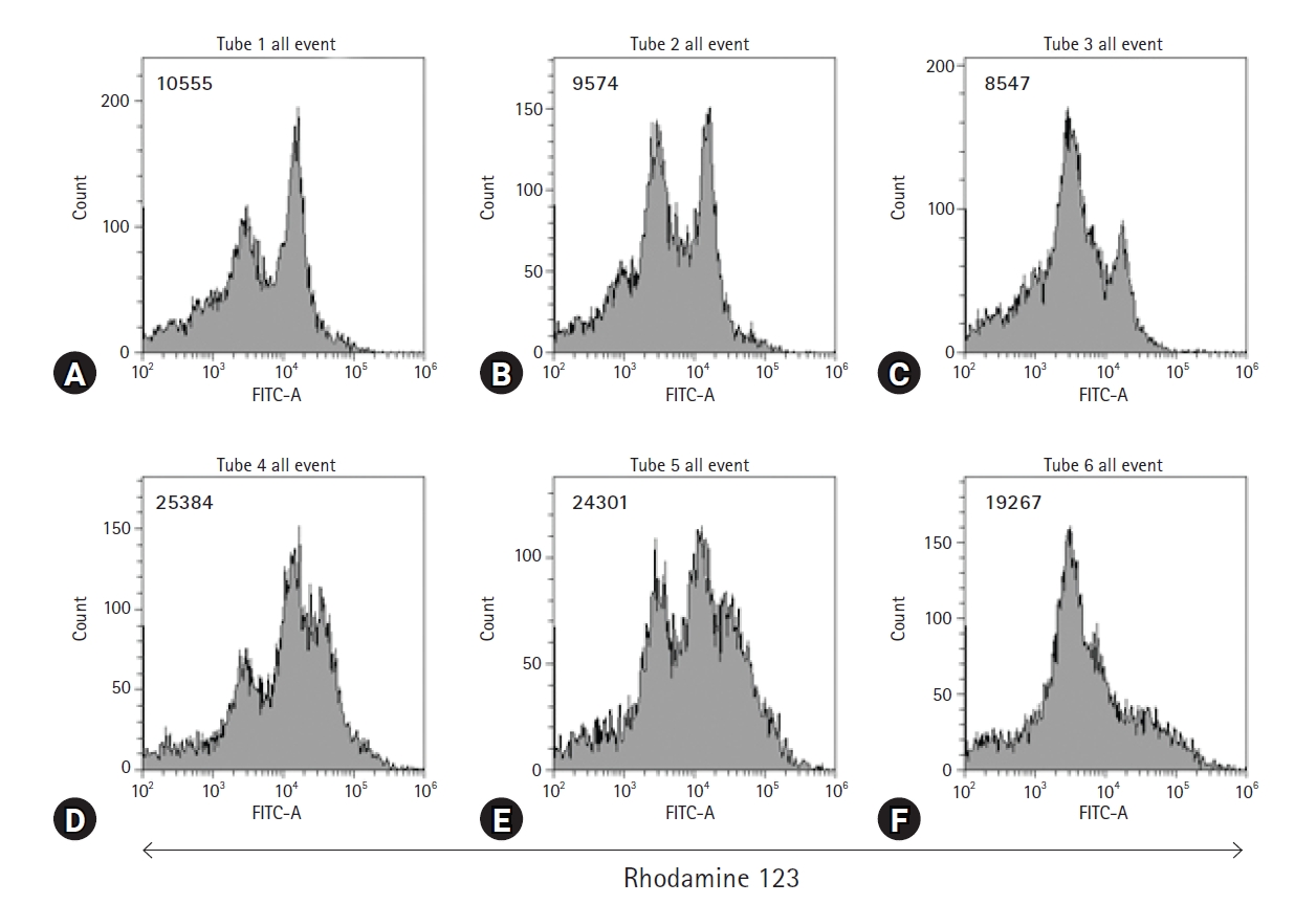
FBZņŚÉ ņØśĒĢ£ Ļ│©ņłśņäĖĒżņØś ļ»ĖĒåĀņĮśļō£ļ”¼ņĢä ļ¦ēņĀäņ£ä ļ│ĆĒÖö
FBZņØ┤ Ļ│©ņłśņäĖĒżņŚÉ ļ»Ėņ╣śļŖö ņśüĒ¢źņØä ņĪ░ņé¼ĒĢśĻĖ░ ņ£äĒĢ┤ FBZĻ│╝ LPSļź╝ ņ▓śļ”¼ĒĢ£ Ļ│©ņłśņäĖĒżņØś ļ»ĖĒåĀņĮśļō£ļ”¼ņĢä ļ¦ēņĀäņ£äļź╝ ņĖĪņĀĢĒĢśņśĆļŗż. ņØ┤ļź╝ ņ£äĒĢ┤ rhodamine 123 ņÜ®ņĢĪņØä ņé¼ņÜ®ĒĢśņŚ¼ Ļ│©ņłśņäĖĒżļź╝ ņŚ╝ņāēĒĢ£ Ēøä ņ£ĀņäĖĒż ļČäņäØņØä ĒĢśņśĆļŗż(Fig. 2). LPSļŖö Ļ│©ņłśņäĖĒżņØś ļ»ĖĒåĀņĮśļō£ļ”¼ņĢä ļ¦ēņĀäņ£äļź╝ ļåÆņØ┤ļŖö ļ░śļ®┤, FBZņØĆ LPSļĪ£ ņ”ØĻ░ĆļÉ£ ļ»ĖĒåĀņĮśļō£ļ”¼ņĢä ļ¦ēņĀäņ£äļź╝ Ēü¼Ļ▓ī Ļ░Éņåīņŗ£ņ╝░ļŗż. ĒŖ╣Ē׳, FBZ 0.25 ┬ĄM ļåŹļÅäņÖĆ FBZ 1 ┬ĄM ļåŹļÅäņØś fluorescence intensity ĒÅēĻĘĀĻ░ÆņØś ņ░©ņØ┤ļŖö 5,034ļĪ£, FBZ 0 ┬ĄM ļåŹļÅäņÖĆ FBZ 0.25 ┬ĄM ļåŹļÅäņØś ĒÅēĻĘĀĻ░ÆņØś ņ░©ņØ┤ņØĖ 1,083ļ│┤ļŗż Ēü¼ļŗż. ņØ┤ Ļ▓░Ļ│╝ļŖö 0.25-1 ┬ĄM ņé¼ņØ┤ ļåŹļÅäņØś FBZņØĆ LPSņŚÉ ņØśĒĢ┤ ņ×ÉĻĘ╣ļÉ£ Ļ│©ņłśņäĖĒżņØś ļ»ĖĒåĀņĮśļō£ļ”¼ņĢä ņØ┤ņżæļ¦ēņØś ĻĄ¼ņĪ░ļź╝ ļŹöņÜ▒ ļČłņĢłņĀĢĒĢśĻ▓ī ĒĢĀ ņłś ņ׳ņØīņØä ļéśĒāĆļéĖļŗż.
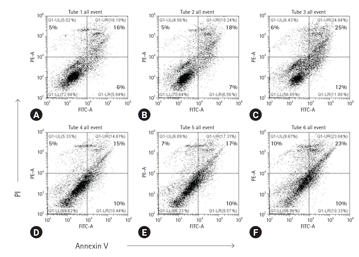
FBZņŚÉ ņØśĒĢ£ Ļ│©ņłśņäĖĒżņØś ņäĖĒżņé¼ ņ”ØĻ░Ć
ņ£ĀņäĖĒż ļČäņäØņØĆ Ļ│©ņłśņäĖĒżņØś ņäĖĒżņé¼ļź╝ ĒÖĢņØĖĒĢśĻĖ░ ņ£äĒĢ┤ annexin V-FITC/PI ņŚ╝ņāē Ēøä ņłśĒ¢ēļÉśņŚłļŗż(Fig. 3). FBZ ļŗ©ļÅģ ļśÉļŖö FBZ + LPS ņ▓śļ”¼ļÉ£ ņäĖĒżņØś viable cells (annexin V-/PI-)ņØś ļ╣äņ£©ņØ┤ ņ£Āņé¼ĒĢśĻ▓ī Ļ░ÉņåīĒĢśņśĆļŗż. ņØ┤ Ļ░ÉņåīļŖö ĒŖ╣Ē׳ Fig. 2ņÖĆ Ļ░ÖņØ┤ FBZ 1 ┬ĄM ļåŹļÅäņŚÉņä£ ĒśäņĀĆĒĢśĻ▓ī Ļ░ÉņåīĒĢ©ņØä ļ│┤ņśĆļŗż. Viable cellsļŖö FBZ ļŗ©ļÅģņØ╝ ļĢī FBZ 0-0.25 ┬ĄMņØĆ 2.3%, FBZ 0.25-1 ┬ĄMņØĆ 13.8% Ļ░ÉņåīĒĢśņśĆļŗż. FBZ + LPS ņ▓śļ”¼ĻĄ░ņŚÉņä£ļÅä FBZ 0-0.25 ┬ĄMņØĆ 3.4%, FBZ 0.25-1 ┬ĄMņØĆ 9.3% Ļ░ÉņåīĒĢśņśĆļŗż. ņŻĮņØĆ ņäĖĒż ņżæ necrotic cells (annexin V-/PI+)ņØś ņłśļŖö FBZ 0-1 ┬ĄM ļåŹļÅäņŚÉņä£ 5%ņŚÉņä£ 6%ļĪ£ ņ”ØĻ░ĆĒĢ£ FBZ ļŗ©ļÅģņŚÉ ļ╣äĒĢ┤ FBZ + LPS ņ▓śļ”¼ĻĄ░ņŚÉņä£ ļŹöņÜ▒ ĒśäņĀĆĒĢ£ ļ│ĆĒÖöļź╝ ļ│┤ņśĆļŗż. ĻĘĖļ¤¼ļéś early apoptosis cells (annexin V+/PI-)ņŚÉņä£ļŖö FBZ ļŗ©ļÅģņØĆ 6%ņŚÉņä£ 12%ļĪ£ ņ”ØĻ░ĆĒĢ£ ļ░śļ®┤, FBZ + LPS ņ▓śļ”¼ĻĄ░ņŚÉņä£ļŖö 10%ļĪ£ ņØ╝ņĀĢĒĢśņśĆļŗż. ņØ┤ļ¤¼ĒĢ£ Ļ▓░Ļ│╝ļŖö FBZņØ┤ LPS ņ▓śļ”¼ ņŚ¼ļČĆņÖĆ Ļ┤ĆĻ│äņŚåņØ┤ Ļ│©ņłśņäĖĒżņŚÉņä£ ņäĖĒżņé¼ļź╝ ņ£ĀļÅäĒĢĀ ņłś ņ׳ņØīņØä ļ│┤ņŚ¼ņżĆļŗż.
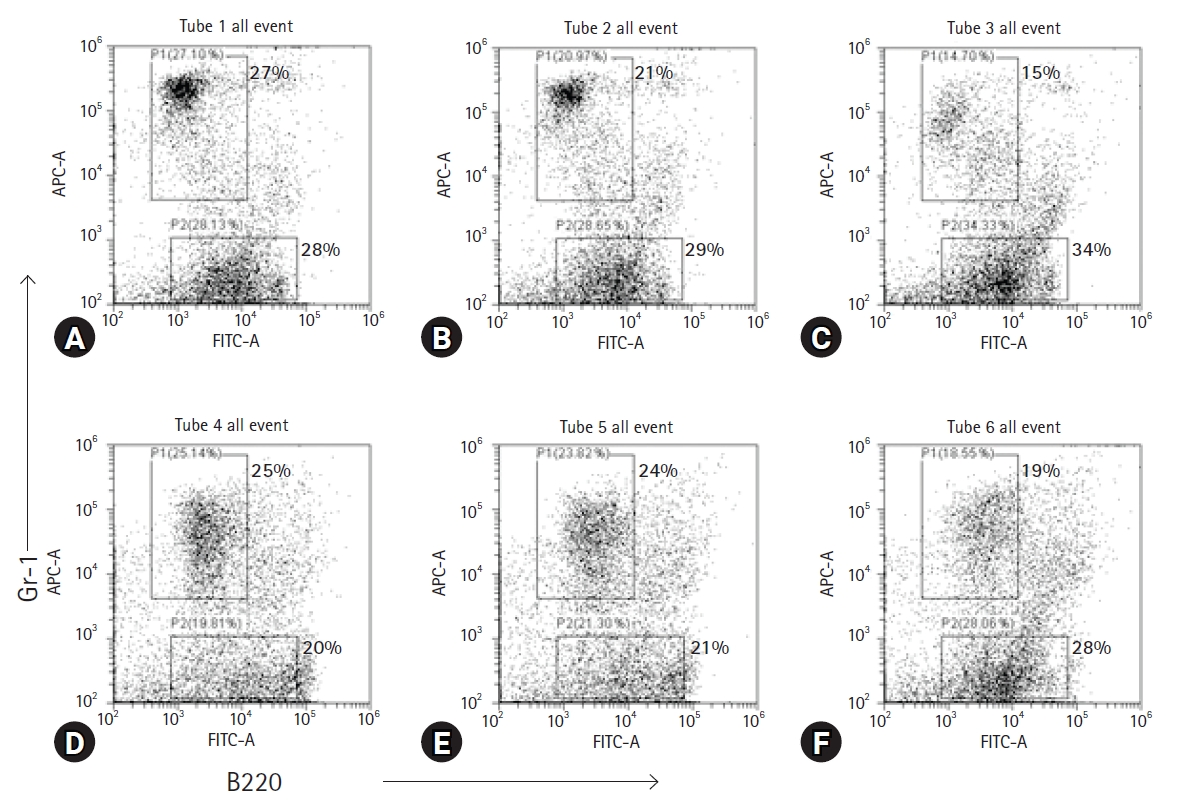
Ļ│©ņłśņäĖĒż ļé┤ņØś Ļ│╝ļ”ĮĻĄ¼ņÖĆ Bļ”╝ĒöäĻĄ¼ņØś ĻĄ¼ņä▒ņŚÉ FBZņØ┤ ļ»Ėņ╣śļŖö ņśüĒ¢ź
ļ¦łņÜ░ņŖż Ļ│©ņłśņäĖĒżņØś ņŻ╝ņÜö ĻĄ¼ņä▒ ņäĖĒż ņżæņŚÉļŖö Ļ│╝ļ”ĮĻĄ¼ņÖĆ Bļ”╝ĒöäĻĄ¼Ļ░Ć ĒżĒĢ©ļÉśņ¢┤ ņ׳ļŗż. ļæÉ Ļ░Ćņ¦Ć ņäĖĒż ĻĄ¼ņä▒ņŚÉ FBZņØ┤ ļ»Ėņ╣śļŖö ņśüĒ¢źņØä ņĪ░ņé¼ĒĢśĻĖ░ ņ£äĒĢ┤, FBZĻ│╝ LPSļĪ£ ņ▓śļ”¼ļÉ£ Ļ│©ņłśņäĖĒżļź╝ Gr-1 ļśÉļŖö B220 ĒŖ╣ņØ┤ņĀüņØĖ ĒĢŁņ▓┤ļĪ£ ņŚ╝ņāēĒĢ£ Ēøä ņ£ĀņäĖĒż ļČäņäØņØä ņŗżņŗ£ĒĢśņśĆļŗż(Fig. 4). Dot plotņØś Gr-1+ ĻĄ¼ņŚŁņØĆ ĒśĖņżæĻĄ¼ļź╝ ĒżĒĢ©ĒĢ£ Ļ│╝ļ”ĮĻĄ¼ņØ┤ļŗż. FBZ 0 ┬ĄM ļåŹļÅäņŚÉņä£ FBZ ļŗ©ļÅģņØĆ 27%, FBZ + LPS ņ▓śļ”¼ĻĄ░ņØĆ 25%ņØĖ Ļ▓āņ£╝ļĪ£ ļ│┤ņĢä LPSļŖö Ļ│╝ļ”ĮĻĄ¼ņØś ļ╣äņ£©ņØä Ēü¼Ļ▓ī ņ”ØĻ░Ćņŗ£Ēéżņ¦Ć ņĢŖņĢśļŗż. ļ░śļ®┤, FBZ 0-1 ┬ĄM ņé¼ņØ┤ņØś ļ│ĆĒÖöļź╝ ļ│┤ļ®┤ FBZ ļŗ©ļÅģņŚÉņä£ļŖö 27%ņŚÉņä£ 15%ļĪ£ Ļ░ÉņåīĒĢśņśĆĻ│Ā FBZ + LPS ņ▓śļ”¼ĻĄ░ņŚÉņä£ļŖö 25%ņŚÉņä£ 19%ļĪ£ Ļ░ÉņåīĒĢśņśĆļŗż. Ļ│©ņłśņäĖĒżņŚÉ LPS ņ▓śļ”¼ņÖĆ Ļ┤ĆĻ│äņŚåņØ┤ Ļ│╝ļ”ĮĻĄ¼ņØś ļ╣äņ£©ņØĆ ĒśäņĀĆĒĢśĻ▓ī Ļ░Éņåīņŗ£ĒéżļŖö ņé¼ņŗżņØä ņĢī ņłś ņ׳ļŗż. Dot plotņØś B220+ ĻĄ¼ņŚŁņØĆ Bļ”╝ĒöäĻĄ¼ņØ┤ļŗż. Bļ”╝ĒöäĻĄ¼ņØś ļ╣äņ£©ņØĆ LPSņŚÉ ņØśĒĢ┤ ņ”ØĻ░ĆĒĢśņ¦Ć ņĢŖņĢśĻ│Ā, 28%ņŚÉņä£ 20%ļĪ£ ņśżĒ׳ļĀż Ļ░ÉņåīĒĢ£ ļ¬©ņŖĄņØä ļ│┤ņśĆļŗż. ļ░śļ®┤, LPS ņ£Āļ¼┤ņÖĆ Ļ┤ĆĻ│äņŚåņØ┤ FBZņØś ļåŹļÅäĻ░Ć ņ”ØĻ░ĆĒĢ©ņŚÉ ļö░ļØ╝ Bļ”╝ĒöäĻĄ¼ņØś ļ╣äņ£©ņØĆ ņ”ØĻ░ĆĒĢśņśĆļŗż.
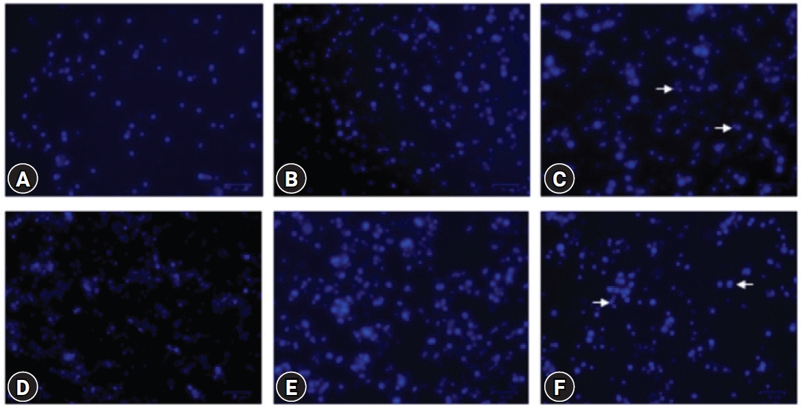
FBZņŚÉ ņØśĒĢ£ Ļ│©ņłśņäĖĒżņØś ĒĢĄ ĒśĢĒā£ ļ│ĆĒÖö
Ļ│©ņłśņäĖĒżļź╝ Hoechst 33342 ņÜ®ņĢĪņ£╝ļĪ£ ņŚ╝ņāēĒĢ£ Ēøä, ĒśĢĻ┤æĒśäļ»ĖĻ▓ĮņØä ņé¼ņÜ®ĒĢśņŚ¼ FBZņŚÉ ņØśĒĢ£ Ļ│©ņłśņäĖĒżņØś ĒĢĄ ĒśĢĒā£ ļ│ĆĒÖöļź╝ Ļ┤Ćņ░░ĒĢśņśĆļŗż(Fig. 5). FBZ ļŗ©ļÅģ ņ▓śļ”¼ĻĄ░ņŚÉņä£ļŖö 1 ┬ĄM ļåŹļÅäņŚÉņä£ ļłłņŚÉ ļØäĻ▓ī ļČäņĀłļÉ£ ĒĢĄņØä Ļ░Ćņ¦ä ņäĖĒżņØś ņłśĻ░Ć ņ”ØĻ░ĆĒĢśņśĆļŗż. LPS ņ▓śļ”¼ļĪ£ Ļ│©ņłśņäĖĒżņØś ņłśĻ░Ć ĒśäņĀĆĒĢśĻ▓ī ņ”ØĻ░ĆļÉśņŚłĻ│Ā FBZņØś 1 ┬ĄM ļåŹļÅäņŚÉņä£ ļČäņĀłļÉ£ ĒĢĄņØä Ļ░Ćņ¦ä ņäĖĒżņØś ņłśĻ░Ć ņ”ØĻ░ĆĒĢśņśĆļŗż. ĒĢĄņØ┤ ļČäņĀłļÉ£ ņäĖĒżĻ░Ć ņ”ØĻ░ĆĒĢśļŖö Ļ▓āņØĆ ņäĖĒżņé¼ņØś ņ”ØĻ░Ćļź╝ ņØśļ»ĖĒĢśļ»ĆļĪ£ FBZ 1 ┬ĄMņØĆ LPS ņ£Āļ¼┤ņÖĆ Ļ┤ĆĻ│äņŚåņØ┤ Ļ│©ņłśņäĖĒżņØś ņäĖĒżņé¼ļź╝ ņ”ØĻ░Ćņŗ£ĒéżļŖö ņé¼ņŗżņØä ĒÖĢņØĖĒ¢łļŗż.
Ļ│Āņ░░
FBZņØĆ ņłśņØśņŚÉņä£ Ļ┤æļ▓öņ£äĒĢśĻ▓ī ņé¼ņÜ®ļÉśļŖö ĒĢŁĻĖ░ņāØņČ®ņĀ£ņĀ£ ņżæ ĒĢśļéśņØ┤ļ®░[1-3], ņĄ£ĻĘ╝ ņóģņ¢æņäĖĒżļź╝ ņØ┤ņÜ®ĒĢ£ ņŚ░ĻĄ¼ņŚÉņä£ ĒĢŁņĢöĒÜ©Ļ│╝ļź╝ Ļ░¢ļŖöļŗżļŖö ņé¼ņŗżņØ┤ ļ░ØĒśĆņĪīļŗż[6]. ĒĢśņ¦Ćļ¦ī FBZņØś ĒĢŁņŚ╝ņ”Ø ĒÜ©Ļ│╝ņŚÉ ļīĆĒĢ£ ņŚ░ĻĄ¼ļŖö ļ¦żņÜ░ ļ»ĖĒØĪĒĢśļ®░ ĻĘĖ ņ×æņÜ®ĻĖ░ņĀäļÅä ņĢīļĀżņĀĖ ņ׳ņ¦Ć ņĢŖļŗż. ļ│Ė ņŚ░ĻĄ¼ņŚÉņä£ļŖö FBZņØś ĒĢŁņŚ╝ņ”Ø ĒÜ©Ļ│╝ļź╝ ņĢīņĢäļ│┤ĻĖ░ ņ£äĒĢ┤ Ļ│©ņłśņäĖĒżņŚÉ ļīĆĒæ£ņĀüņØĖ ņŚ╝ņ”Øņä▒ ņ£ĀļÅäļ¼╝ņ¦ł ņżæ ĒĢśļéśņØĖ LPSļź╝ FBZĻ│╝ ĒĢ©Ļ╗ś ļ│ĄĒĢ® ņ▓śļ”¼ĒĢ£ Ēøä ļČäņäØĒĢśņśĆļŗż.
FBZņØ┤ ņäĖĒżĒÖ£ņä▒ļÅäņÖĆ ļ»ĖĒåĀņĮśļō£ļ”¼ņĢä ļ¦ēņĀäņ£äļź╝ Ļ░Éņåīņŗ£ņ╝£ Ļ│©ņłśņäĖĒżņØś ĒÖ£ņä▒ņØä ņ¢ĄņĀ£ĒĢśņśĆļŗż. ļśÉĒĢ£, annexin V-FITC/PI ņŚ╝ņāēĻ│╝ Hoechst 33342 ņŚ╝ņāēņØä ĒåĄĒĢ┤ FBZņØ┤ ņŚ╝ņ”Øņä▒ Ļ│©ņłśņäĖĒżņØś ņäĖĒżņé¼ļź╝ ņ£ĀļÅäĒĢśļŖö ņé¼ņŗżņØä ĒÖĢņØĖĒĢśņśĆļŗż. ņØ┤ļ¤¼ĒĢ£ Ļ▓░Ļ│╝ļŖö LPS ņ▓śļ”¼ļĪ£ ļ¦īļōżņ¢┤ņ¦ä ņŚ╝ņ”Øņä▒ Ļ│©ņłśņäĖĒżņŚÉņä£ FBZņØ┤ ņäĖĒż ņłśņÖĆ ļ»ĖĒåĀņĮśļō£ļ”¼ņĢä ĻĖ░ļŖźņØä ļ¬©ļæÉ Ļ░Éņåīņŗ£Ēé©ļŗżļŖö Ļ▓āņØä ļ│┤ņŚ¼ņżĆļŗż. Ļ│╝ļ”ĮĻĄ¼ņÖĆ Bļ”╝ĒöäĻĄ¼ņØś ĻĄ¼ņä▒ ļ╣äņ£© ļ│ĆĒÖöļź╝ ļČäņäØĒĢ£ Ļ▓░Ļ│╝ FBZņØĆ LPSĻ░Ć ņ▓śļ”¼ļÉ£ Ļ│©ņłśņäĖĒżņŚÉņä£ Bļ”╝ĒöäĻĄ¼ļ│┤ļŗż Ļ│╝ļ”ĮĻĄ¼ņØś ļ╣äņ£©ņØä ļŹö Ļ░Éņåīņŗ£ņ╝░ļŗż.
FBZņØ┤ Ļ│╝ļ”ĮĻĄ¼ņØś ļ░£ĒśäņØä ņäĀĒāØņĀüņ£╝ļĪ£ ņ¢ĄņĀ£ĒĢśļŖö ņĀĢĒÖĢĒĢ£ ĻĖ░ņĀäņØĆ ļ░ØĒśĆņ¦Ćņ¦Ć ņĢŖņĢśņ¦Ćļ¦ī, ņØ┤ļź╝ ņäżļ¬ģĒĢĀ ņłś ņ׳ļŖö ņäĀĒ¢ēņŚ░ĻĄ¼ļōżņØ┤ ņ׳ļŗż. ņ▓£ņŗØ ļÅÖļ¼╝ļ¬©ļŹĖņŚÉņä£ FBZņØ┤ ņĢīļĀłļź┤ĻĖ░ņä▒ ĻĖ░ļÅäņŚ╝ņ”ØĻ│╝ Th2 ņé¼ņØ┤ĒåĀņ╣┤ņØĖņØś ņāØņé░ņØä Ļ░Éņåīņŗ£ņ╝░ļŗż[12]. FBZņØĆ ĻĖ░ņāØņČ® Ļ░ÉņŚ╝ņ”Ø ņ╣śļŻīņŚÉ ļŗżņ¢æĒĢśĻ▓ī ņØ┤ņÜ®ļÉ£ļŗż[13]. ņØ╝ļ░śņĀüņ£╝ļĪ£ ĻĖ░ņāØņČ® Ļ░ÉņŚ╝ ņŗ£ ĒśĖņé░ĻĄ¼ļź╝ ĒżĒĢ©ĒĢ£ Ļ│╝ļ”ĮĻĄ¼Ļ░Ć ņ”ØĻ░ĆļÉśĻ│Ā[14], FBZņØ┤ ĻĖ░ņāØņČ® ņ╣śļŻīņŚÉ ņØ┤ņÜ®ļÉśļ»ĆļĪ£, FBZņØ┤ Ļ│╝ļ”ĮĻĄ¼ņØś ņł½ņ×É ļśÉļŖö ĻĖ░ļŖźņŚÉ ņśüĒ¢źņØä ļ»Ėņ╣Ā ņłś ņ׳ņØīņØä ņ£ĀņČöĒĢĀ ņłś ņ׳ļŗż. ļśÉĒĢ£, squirrel monkeyļź╝ ņØ┤ņÜ®ĒĢ£ ņĢ×ņäĀ ņŚ░ĻĄ¼ņŚÉņä£ ņ┤Ø ļ░▒ĒśłĻĄ¼, ĒśĖņżæĻĄ¼, ļŗ©ĒĢĄĻĄ¼ ļ░Å ĒśĖņé░ĻĄ¼ ņłś ļ╣äĻĄÉļŖö FBZņØä ņ▓śļ”¼ĒĢśņ¦Ć ņĢŖņØĆ ļīĆņĪ░ĻĄ░ņŚÉ ļ╣äĒĢ┤ FBZņØä ņ▓śļ”¼ĒĢ£ ĻĄ░ņŚÉņä£ ĒśäņĀĆĒ׳ Ļ░ÉņåīĒĢ£ ļ¬©ņŖĄņØä ļ│┤ņśĆļŗż[15]. ņØ┤ļź╝ ņóģĒĢ®ĒĢśļ®┤ FBZņØĆ Ļ│©ņłśņäĖĒż ņżæņŚÉņä£ļÅä Ļ│╝ļ”ĮĻĄ¼ļź╝ ĒŖ╣Ē׳ Ļ░Éņåīņŗ£Ēé©ļŗżĻ│Ā ņāØĻ░üļÉ£ļŗż. ļ░śļ®┤, FBZņØś ļåŹļÅäĻ░Ć ņ”ØĻ░ĆĒĢ©ņŚÉ ļö░ļØ╝ ņĀäņ▓┤ ņāØņĪ┤ ņäĖĒż ņłśņÖĆ Ļ│╝ļ”ĮĻĄ¼ļŖö ņżäņ¢┤ļō£ļŖöļŹ░, ļ”╝ĒöäĻĄ¼ņØś ņĀłļīĆĻ░ÆņØĆ Ēü¼Ļ▓ī ļ│ĆĒĢśņ¦Ć ņĢŖņĢä ņāüļīĆņĀüņØĖ ļ╣äņ£©ļĪ£ ļ”╝ĒöäĻĄ¼Ļ░Ć ņ”ØĻ░ĆĒĢ£ļŗżĻ│Ā ņČöņĖĪĒĢĀ ņłś ņ׳ļŗż. ļŗżļźĖ ņŚ░ĻĄ¼ņŚÉņä£ļÅä ņĀłļīĆ ļ”╝ĒöäĻĄ¼ ņłśļŖö FBZ ņ╣śļŻīņŚÉ ņśüĒ¢źņØä Ļ▒░ņØś ļ░øņ¦Ć ņĢŖņĢśļŗż[15]. ĒĢśņ¦Ćļ¦ī FBZņØĆ ļ¦łņÜ░ņŖżņØś ļ®┤ņŚŁņ▓┤Ļ│äņŚÉ ņāüļŗ╣ĒĢ£ ņśüĒ¢źņØä ļ»Ėņ╣śļŖö Ļ▓āņ£╝ļĪ£ ļ│┤ņĢä[16], Bļ”╝ĒöäĻĄ¼ņØś ĻĖ░ļŖźņØ┤ Ļ░ÉņåīļÉĀ Ļ░ĆļŖźņä▒ņØĆ ņ׳ļŗż.
ļŗżļźĖ ņŚ░ĻĄ¼ņŚÉņä£ļŖö ņäżņ╣śļźśņŚÉņä£ FBZņŚÉ ņØśĒĢ£ Ļ│©ņłś ņ¢ĄņĀ£Ļ░Ć ļ░£ņāØĒĢśņ¦Ć ņĢŖņØīņØä ļ│┤Ļ│ĀĒĢśņśĆļŗż[17]. ĒĢśņ¦Ćļ¦ī ļ│Ė ņŚ░ĻĄ¼ņŚÉņä£ļŖö Ļ│©ņłśņäĖĒżņŚÉ ļīĆĒĢ£ FBZņØś ņ¦üņĀæņĀüņØĖ ĒÜ©Ļ│╝ļź╝ ņĖĪņĀĢĒĢśņŚ¼ ņŗżĒŚśņĪ░Ļ▒┤ņØ┤ ļŗżļź┤ļŗż. ļśÉĒĢ£ LPSņŚÉ ņØśĒĢ┤ Ļ│╝ļÅäĒĢ£ ņŚ╝ņ”Ø ļ░śņØæņØ┤ ļ░£ņāØĒĢ£ Ļ│©ņłśņäĖĒżļź╝ ņØ┤ņÜ®ĒĢśņŚ¼ FBZņØś ĒĢŁņŚ╝ņ”Ø ĒÜ©Ļ│╝ņŚÉ ļīĆĒĢ£ ņŚ░ĻĄ¼ļź╝ ņłśĒ¢ēĒ¢łņ£╝ļ®░, FBZņØä ĒĢŁņŚ╝ņ”ØņĀ£ļĪ£ ņé¼ņÜ®ĒĢĀ ņłś ņ׳ļŖö Ļ░ĆļŖźņä▒ņØä ĒÖĢņØĖĒ¢łļŗż.
ņØ┤ļ¤¼ĒĢ£ Ļ▓░Ļ│╝ļź╝ ņóģĒĢ®ĒĢśļ®┤, LPS ņ▓śļ”¼ļĪ£ ņØĖĒĢ┤ ņŚ╝ņ”Øņä▒ ņāüĒā£Ļ░Ć ļÉ£ Ļ│©ņłśņäĖĒżņŚÉņä£ FBZņØĆ ļ»ĖĒåĀņĮśļō£ļ”¼ņĢä ļ¦ēņĀäņ£äļź╝ ļČłņĢłņĀĢĒĢśĻ▓ī ļ¦īļōżņ¢┤ ņäĖĒżņé¼ļź╝ ņ£ĀļÅäņŗ£ņ╝£ ĒĢŁņŚ╝ņ”Ø ĒÜ©Ļ│╝ļź╝ ņØ╝ņ£╝ĒéżļŖö Ļ▓āņ£╝ļĪ£ ņČöņĀĢļÉ£ļŗż. Ē¢źĒøä ļ»ĖĒåĀņĮśļō£ļ”¼ņĢäņÖĆ FBZņØś Ļ┤ĆļĀ©ņä▒, ļŗżļźĖ ĒĢŁņŚ╝ņ”ØņĀ£ņÖĆņØś ņāüņŖ╣ ĒÜ©Ļ│╝, ļŗżņ¢æĒĢ£ ņŚ╝ņ”ØņØ┤ ņ£ĀļÅäļÉ£ ļÅÖļ¼╝ņ¦łĒÖś ļ¬©ļŹĖņŚÉņä£ņØś ņĢĮĒÜ© ņŗżĒŚśņØ┤ ņČöĻ░ĆņĀüņ£╝ļĪ£ ĒĢäņÜöĒĢśļŗż.
 PDF Links
 PubReader
 ePub Link
 Full text via DOI
 Download Citation
 Print
Print


