Salmonella enterica serotype Choleraesuis is a facultative intracellular pathogen that causes swine paratyphoid, with clinical manifestations of enterocolitis and septicemia [
1]. Infection with the septicemic
S. Choleraesuis is considered a differential diagnosis for pigs with African swine fever virus (ASFV), since it shows similar clinical signs and lesions [
2].
In the 1950s and 1960s,
S. Choleraesuis was the predominant serotype in the global pig industry [
1]. However, it is at present rare in the European Union and Australia [
1,
3]. As
S. Choleraesuis has diminished,
Salmonella Typhimurium has become the most frequently isolated serotype [
4]. Despite its low prevalence in pigs,
S. Choleraesuis is becoming more prevalent in wild boars from Europe, which are suspected to be carriers [
1,
5]. Furthermore, antimicrobial resistance to
S. Choleraesuis has been reported in many countries [
1,
2,
6]. To control the occurrence of salmonellosis in pigs, antimicrobial administration to the affected animals is necessary, and antimicrobial susceptibility testing is required for the selection of empirical antimicrobials [
5].
In Korea, there are many studies on salmonellosis in pigs; however, most are focused on S. Typhimurium, the most common serotype in Korea [
6,
7]. Although two isolates of
S. Choleraesuis were isolated from diarrheic pigs in Korea in 2004, information on
S. Choleraesuis in Korea is limited [
8]. Herein, we describe the clinical signs, pathological lesions, and laboratory results of
S. Choleraesuis infections in weaned pigs in Korea.
In August 2020, a farrow-to-finish pig farm in Yangsan, Republic of Korea, breeding 2,000 pigs, reported lethargy and sudden death of weaned pigs (40 days old) that exhibited mucoid diarrhea and cough. Twenty-five percent (100/400) of the weaned pigs exhibited clinical signs, and 20 pigs died (mortality rate, 5%). Two weaned pigs were referred to the Animal and Plant Quarantine Agency for necropsy and differential diagnosis.
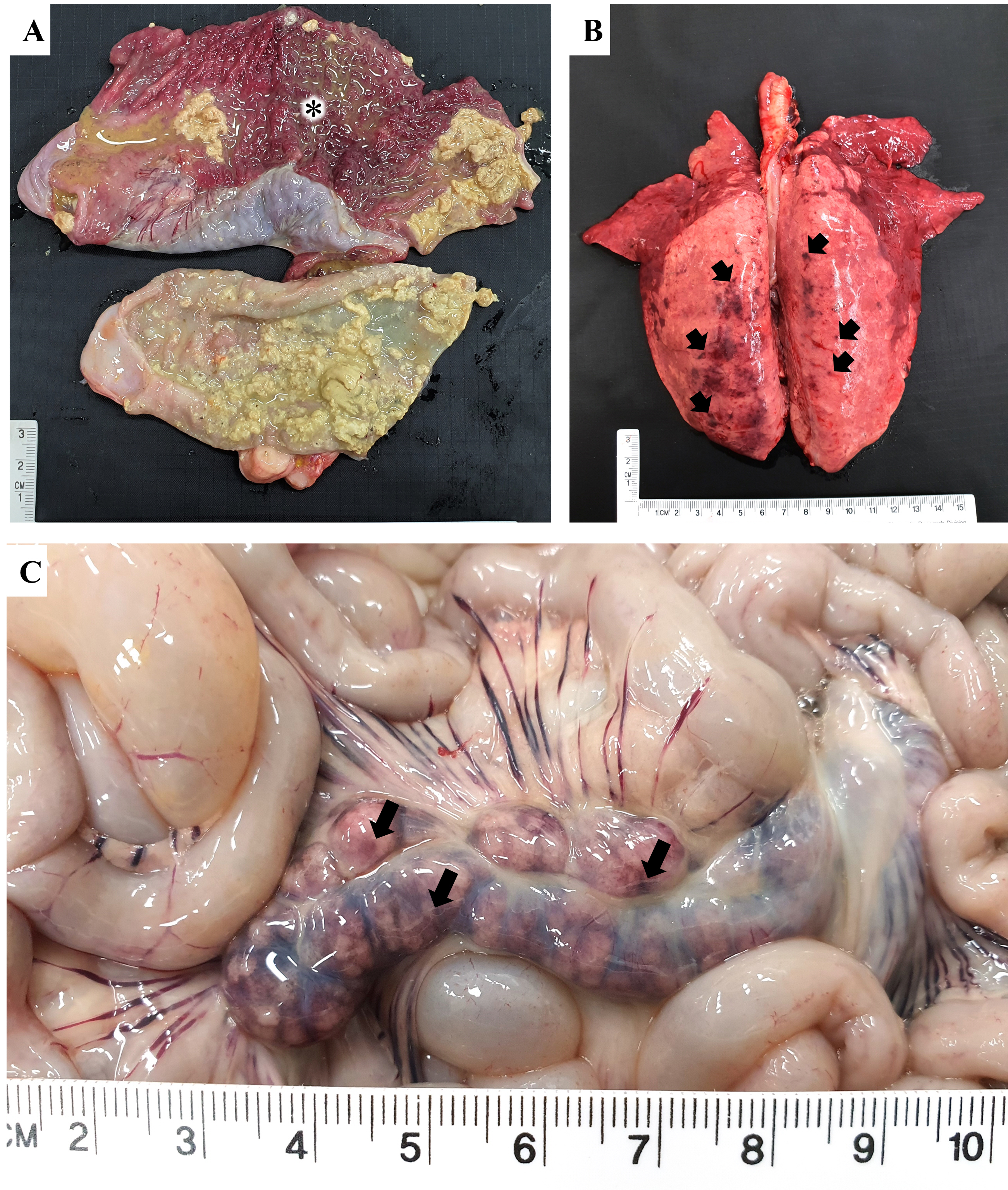
Both pigs exhibited non-collapsed lungs with consolidation. Pig 1 had severe hyperemic mucosa in the colon, with a yellow, fibrinous membrane and pasty contents (
Fig. 1A). Pig 2 had consolidation in the cranioventral lobes and multifocal ecchymoses in the dorsocaudal lobes of the lungs (
Fig. 1B). Furthermore, the mesenteric lymph nodes were enlarged and congested (
Fig. 1C).
After necropsy, representative tissues, including the brain, lungs, liver, spleen, kidneys, small intestines, large intestines, and lymph nodes, were fixed in 10% neutral buffered formalin for 24 hours. The fixed tissues were processed according to a previous study [
9], and 2-┬Ąm sections were stained with hematoxylin and eosin. Histopathologically, both pigs exhibited severe bronchointerstitial pneumonia (
Fig. 2A). In pig 1, severe necrosis of the intestinal epithelial cells and cryptic dilation were observed, and submucosal infiltration of macrophages and lymphocytes were also detected in the colon (
Fig. 2B). In pig 2, moderate perivascular infiltration of mononuclear cells with bacterial colonies and multifocal gliosis were observed in the cerebrum (
Fig. 2C and
D). Lesions corresponding to hemorrhages, lympholysis, and bacterial colonization were observed in the spleen and mesenteric lymph nodes of pig 2 (
Fig 2E-G). In addition, coagulative necrosis and bacterial colonies were observed in the liver of pig 2 (
Fig. 2H).
The lungs and small and large intestinal contents with their gross lesions were aseptically collected and cultured onto sheep blood agar (Asan Pharmaceutical Co. Ltd., Korea) and MacConkey agar (Becton; Dickinson and Company, USA) under 5% CO
2 at 37Ōäā for 24 hours. The identification of
S. Choleraesuis was confirmed using the AccuPower
Salmonella spp. 3-plex polymerase chain reaction (PCR) Kit (Bioneer Corporation, Korea), according to the manufacturerŌĆÖs instructions. For the detection of major porcine viruses, including the porcine reproductive and respiratory syndrome virus (PRRSV), the classical swine fever virus (CSFV), the swine influenza virus (SIV), the porcine circovirus type 2 (PCV2), the transmissible gastroenteritis virus (TGEV), the porcine epidemic diarrhea virus (PEDV), and rotaviruses, we used the VDx PRRSV HPMP RT-PCR, CSFV 5ŌĆÖNCR RT-PCR, SIV RT-PCR, and PCV2 qPCR kits (Median Diagnostics Inc., Korea), the LiliF TGEV/PEDV RT-PCR kit (iNtRON Biotechnology Inc., Korea), and the POBGEN Rotavirus (A, B, C) detection kit (POSTBIO Inc., Korea), according to the manufacturerŌĆÖs instructions. In addition, serotyping procedures were performed as previously described [
6]. Antimicrobial susceptibility testing was performed with the disc diffusion method according to a previous study [
1]. The following discs (Oxoid Ltd., UK) were used: ampicillin (10 ┬Ąg), ceftiofur (30 ┬Ąg), enrofloxacin (5 ┬Ąg), gentamicin (10 ┬Ąg), and tetracycline (30 ┬Ąg).
Escherichia coli ATCC 25922 was used as a control strain.
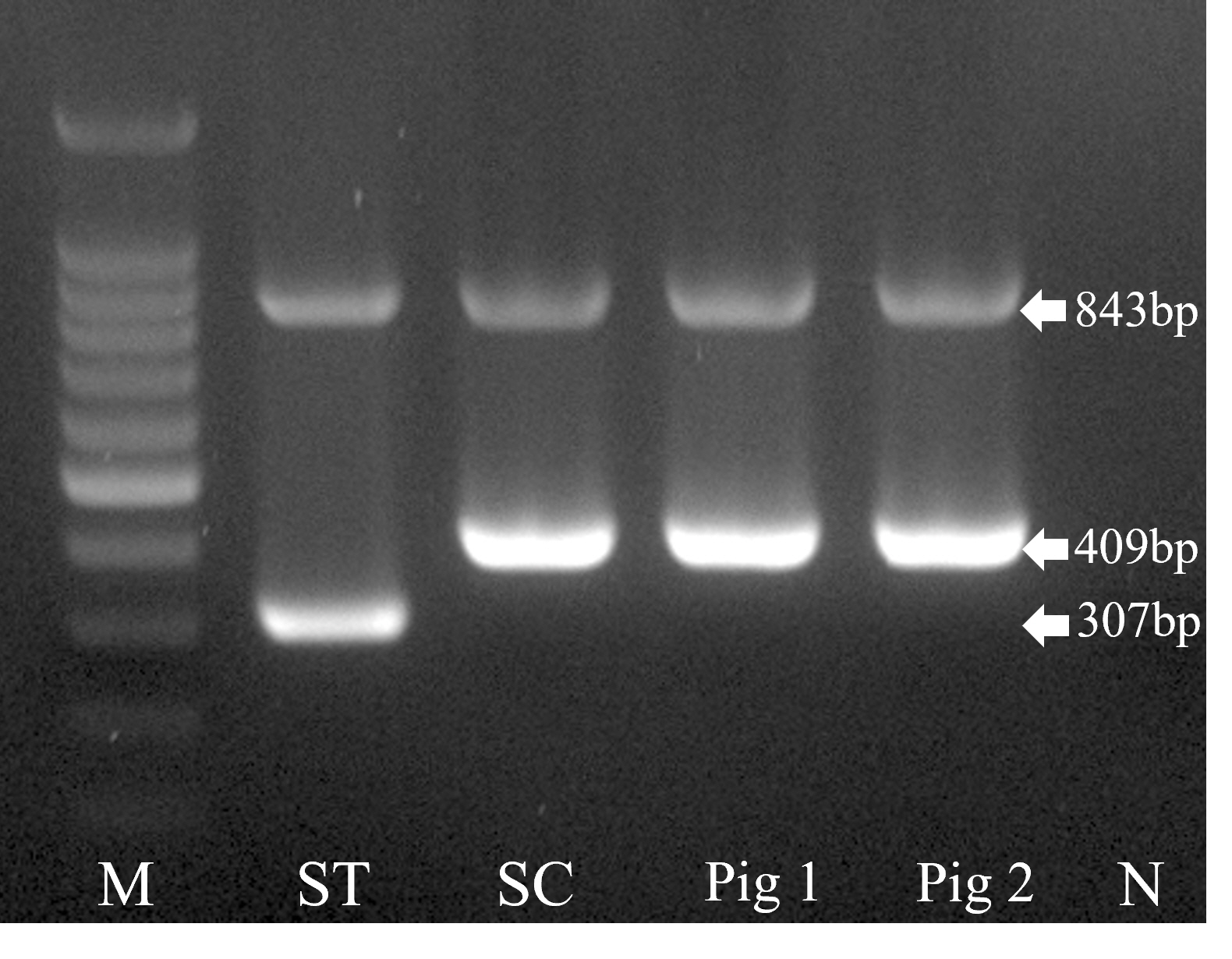
Salmonella-suspected colonies were isolated from the large intestinal contents (pig 1), lungs (pig 2), and small intestinal contents (pig 2). Based on the serotyping procedures, all
Salmonella spp. isolates belonged to the C1 group, serovar 6,7:c:1,5, and were confirmed as
S. Choleraesuis via PCR (
Fig. 3). In addition,
Streptococcus suis was isolated from the lungs of pig 1. PRRSV was detected in all the lung samples of both pigs. The isolates from lungs and intestines exhibited resistance to multiple antimicrobials (ampicillin, gentamicin, and tetracycline) according to the results of the antimicrobial susceptibility test.
Septicemic salmonellosis caused by
S. Choleraesuis has rarely been reported in domestic pigs [
3]. In this study, histopathological lesions were discovered in the large intestines of pig 1, and in the systemic organs (i.e., the brain, lungs, liver, spleen, and mesenteric lymph nodes) of pig 2. Although
S. suis was isolated from the lungs of pig 1, we believe that this common swine pulmonary isolate was associated only with the bronchopneumonia [
10]. Based on the histopathological and laboratory examinations, pigs 1 and 2 were diagnosed with enterocolitis and septicemia, respectively, both caused by
S. Choleraesuis. A previous study suggested that PRRSV may increase the susceptibility of pigs to
S. Choleraesuis infections [
11]. Synergistic interactions between PRRSV and
S. Choleraesuis may explain the relatively high incidence rate (25%), mortality rate (5%), and systemic pathological lesions among pigs on the farm in this study [
11]. The overlap in gross lesions caused by these microbes (PRRSV,
S. Choleraesuis, and
S. suis) complicates the diagnosis, and the pathogenic significance of
S. Choleraesuis isolates in pigs needs to be elucidated through experimental animal studies.
S. Choleraesuis isolates from pigs and wild boars are known to display resistance to different varieties of antimicrobials [
1,
12]. In this study, all of the
S. Choleraesuis isolates also exhibited resistance to the three antimicrobials most frequently used in pigs in Korea (ampicillin, gentamicin, and tetracycline) [
13]. Further antimicrobial characterization studies with minimum inhibitory concentrations for a variety of antimicrobials is required to elucidate the antimicrobial resistance pattern of the
S. Choleraesuis isolates.
African swine fever has been reported in domestic and wild pigs in Korea since September 2019 and remains a substantial threat to the pig industry [
14]. Septicemic salmonellosis causes specific gross lesions, such as splenomegaly and congestive swelling in the lymph nodes, similar to ASFV infection [
2]. However, the only gross lesions found in the current study were enlarged and congested mesenteric lymph nodes. On the other hand, all the pigs on the farm were administered antimicrobials (colistin sulfate) by a veterinary practitioner after the onset of the clinical signs. These treatment attempts reportedly result in milder disease in cases of
S. Choleraesuis infection [
2].
Recently, the occurrences and characteristics of
S. Choleraesuis in wild boars have been reported in Europe, suggesting the necessity of
S. Choleraesuis surveillance in wildlife [
1,
5]. To the best of our knowledge, there is limited information on
S. Choleraesuis isolates in Korea, and this is the first clinicopathological report of such infections in domestic pigs in Korea. Future surveillance investigations of
S. Choleraesuis infections in domestic and wild pigs are required, and vaccines need to be considered to prevent a severe outbreak of this pathogen in Korea.


 PDF Links
 PubReader
 ePub Link
 Full text via DOI
 Download Citation
 Print
Print


