Hydramnios related to fetal deformity in a Hanwoo cow: a case report
Article information

Abstract
A Hanwoo cow with a delayed gestation and abdominal distension was delivered following PGF2α injection. There was excessive amniotic fluid, and a male calf was delivered but died immediately. The calf had no eyes and nose, and a cleft palate on the upper jaw. Gross appearance and computed tomography image showed that upper teeth were spread out on both sides due to cleft palate in the upper jaw, and lower jaw and teeth were positioned upward. There were no other brain parts except cerebellum. These findings show a rare case of hydramnios related to fetal congenital deformity in a Hanwoo cow.
Hydrops of the amnion (hydramnios) is a condition in which excessive amniotic fluid is gradually accumulated in the amniotic sac during the last trimester of pregnancy and occurs in approximately 10% of the cases of hydrops of the fetal membranes in cows [1]. Hydramnios is caused by an abnormal fetus that prevents fetal swallowing or intestinal transport of amniotic fluid, whereas hydrallantosis is due to uterine abnormalities (abnormal placentation) of the dam [2]. The incidence of hydramnios is rare and only few cases have been reported in buffalos [3,4] and cattle [5], however this condition has never been reported in Hanwoo cows. During late gestation, the volume of normal amniotic fluid reaches between 3.8 and 7.6 L, however, the volume may increase to 19 to 114 L in hydramnios [1]. The cow with hydramnois does not show overt clinical signs due to gradual increase of amniotic fluid, and the prognosis for the future reproductive performance of the dam is generally good, whereas the fetus is invariably defective and fatal [6].
Fetal deformity is defined as a morphogenesis abnormality that occurs during intrauterine life and is observed at birth [7]. It can develop at any point during pregnancy and is thought to be caused by a variety of reasons, including genetics and environmental factors. It is known that ACAN PRKG2, EVC2, MOCS1, CLCN7, SLC4A2, FBN1, SLC35A3, FANC1, and LRP4/Mefg7 gene mutations influence various types of congenital skeletal deformities [8]. Toxic plants such as Veratrum californicum, Wild lupins, Leucaena, and Mimosa, vitamin A deficiency, viral infection including Schmallenberg virus, Akabane virus and bunyaviruses, and parbendazole trigger congenital skeletal deformities in an environmental way [8]. Most representative congenital skeletal deformities are cleft palate, brachygnatia achondroplasia, congenital paunch calf syndrome, complex vertebral malformation, arachnomelia, short spine lethal, perosomun acaudatus, and syndactyly [9].
The present case reports an example of hydramnios resulted from a fetal congenital deformity in a primiparous Hanwoo cow for the first time.
The expected delivery date for a 2 year-old primiparous Hanwoo cow, which weighed about 400 kg and maintained on a Hanwoo farm in Chungcheong Province, was scheduled on February 24, 2022. There were no overt signs of delivery until March 7, 2022: there was no obvious udder enlargement and no evidence of straining, although the abdomen was distended excessively as in case of twins-pregnancy. Thus, the induction of parturition was induced with an injection of 30 mg of PGF2α, dinoprost tromethamine (Lutalyse; Zoetis, Belgium) on March 7, 2022. Consequently, on March 10, 2022, the cow gave birth to a calf following gentle traction. At delivery, viscous and syrupy amniotic fluid was much more than as usual (approximately 40 L) and the calf presented in the posterior position in birth canal. The delivered calf was alive for a few minutes but died immediately. The sex of calf was male, and the body weight was 20 kg.
As for the calf’s characteristic appearance, there were no eyes and nose, and there was cleft palate on the upper jaw (Fig. 1). The characteristic autopsy findings were as follows: the cranial cavity was very small and there were no other parts in the cavity except the cerebellum (Fig. 2A), which was connected to the spinal cord (Fig. 2B).
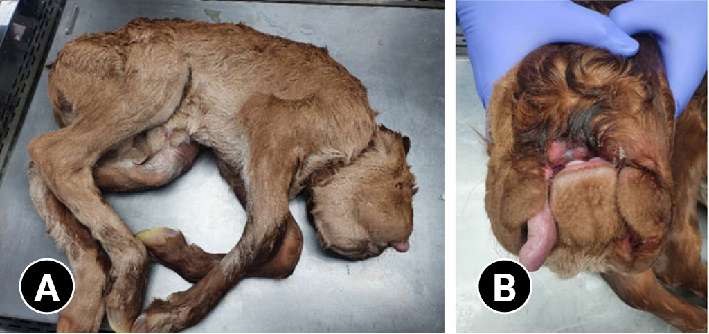
Gross appearance of (A) a full-body calf after delivery, showing (B) a state of extreme deformity of head portion.

Gross findings of cranial part, which shows (A) only the cerebellum in cranial cavity (surrounded by arrows). (B) Brain shows only cerebellum (a) and spinal cord (b).
The scan was performed using a 4-slice computed tomography (CT) scanner (Hi Speed Qx/I; GE Medical Co., USA). CT images were acquired using a helical CT unit. The slice thickness and interval in patient was 1.25 mm with a pitch of 1.5, using the following parameter: 100 mAS tube, 120 kV tube voltage, 500 mm field of view, 512 × 512 matrix. CT images were reviewed using DICOM viewing software (RadiAnt DICOM viewer; Medixant, Poland). CT images were examined by 3-dimensional models and multiplanar reconstruction images. Fig. 3 show gross appearance and CT image of facial part of the calf, respectively. Due to cleft palate in the upper jaw, the upper teeth were spread out on both sides of the face, and the lower jaw and teeth were positioned upward. Except edema in the adrenal gland and incomplete hepatic lobe differentiation, no other morphological abnormalities were observed in other organs. Likewise, autopsy findings, together with CT image showed fetal deformities, especially severe abnormalities in the head.
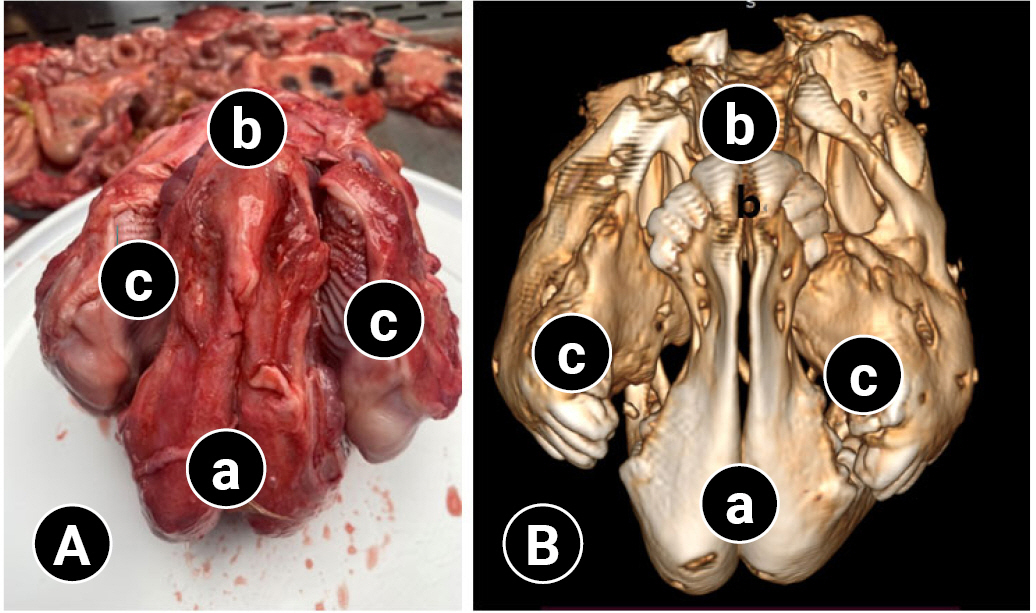
(A) Gross appearance of head part during autopsy. (a) mandible; (b) lower teeth covered membrane, positioned upward; (c) upper teeth, spread out on both sides of the face. (B) Computed tomography image of the facial part of the calf. (a) mandible; (b) lower teeth, positioned upward; (c) upper teeth, spread out on both sides of the face.
After the necropsy, all organ tissues were collected, and histopathologic analysis was conducted. Briefly, all specimens were fixed in 10% neutral buffered formalin and processed routinely. The paraffin blocks were sectioned to 5 μm thickness and arranged on silane-coated slides. Sections were stained with hematoxylin and eosin.
Histologically, multiple organ congestion and hemorrhage were observed. Mucosal and submucosal layers were destroyed with considerable bleeding in the abomasum (Supplementary Fig. 1A), and mild hemorrhage was seen in the villi of the large intestine (Supplementary Fig. 1B). The kidney (Supplementary Fig. 1C) and lung (Supplementary Fig. 1D) also showed severe and diffuse congestion and hemorrhage. Furthermore, diffuse hemorrhage in the cerebellum was seen most prominently in white matter (Supplementary Fig. 1E). Multiple organ dysfunction and hemorrhage can occur in the asphyxiated condition in neonates [10], and in our case, multiple organ hemorrhage appears to be caused by sudden respiratory failure due to congenital cranial anatomical anomalies. Diffuse cerebellar meningitis (Supplementary Fig. 2A) and pulmonary edema (Supplementary Fig. 2B) were also found, both of which are thought to be induced by asphyxia [11]. In adrenal glands, a unilateral cyst with a diameter of 0.8 cm consisted of epithelial lining between the adrenal capsule and cortex (Supplementary Fig. 3B), and mild adrenocortical hyperplasia (Supplementary Fig. 3D) was observed compared to the normal adrenal gland on the opposite side (Supplementary Fig. 3A and C). It is unlikely to be a functioning adrenal cyst, since functioning adrenal cysts are usually associated with tumors.
The present case reports a primiparous Hanwoo cow with a delayed gestation, as diagnosed of having hydramnios: the cow’s abdomen was extended as in twins-pregnancy due to excessive amniotic fluid accumulation, which may be related to fetal deformities, mainly concentrated in the head part (aplasia of parts of the brain other than the cerebellum, eyes, and nose, and presence of cleft palate on the upper jaw).
There was a prolonged gestation (~11 days) in the presented Hanwoo cow, which seems to be related to fetal congenital deformities, especially absence of pituitary gland, by which adrenocorticotropic hormone and cortisol were not secreted from the fetus. It is known that the trigger for onset of parturition is an increased cortisol production by the adrenal gland of fetus [12]. Similar abnormalities include pituitary hypoplasia in Guernsey cattle and bulldog calves in Dexters [13]. In addition, it was reported that a buffalo diagnosed of having both hydramnios and hydrallantois had no brain tissue in the cranial cavity [4]. The other visible feature in the present case was that there was distinct abdominal distension, as seen in the twins-pregnancy [13], originated from excessive accumulation of amniotic fluid in the amniotic sac. This condition may be associated with coincidental oral deformities, especially cleft palate, which might prevent the calf from swallowing amniotic fluid [4,13]. The calf’s death immediately after delivery, while alive for a few minutes, was also associated with the fetal head deformities without nose, which made breathing impossible after the navel was detached.
The present case reports a rare case of hydramnios in a primiparous Hanwoo cow, that had an extended gestation (~11 days) and abdominal distension due to excessive accumulation of amniotic fluid, with distinct fetal deformities, mainly concentrated in the head part (absence of parts of the brain other than the cerebellum, eyes, and nose, and presence of cleft palate on the upper jaw).
Supplementary Materials
Supplementary data are available at https://doi.org/10.14405/kjvr.20220029.
Histopathologic images of hemorrhage in multiple organs. Congestion and hemorrhage were observed in multiple organs including (A) abomasum, (B) large intestine, (C) kidney, (D) lung, and (E) cerebellum under a microscope. H&E, scale bars: (A, B) 500 µm, (C-E) 2 mm.
Histopathologic changes in the cerebellum and lung tissue. (A) Mononuclear inflammatory cell infiltration in cerebellar meninges (arrows) and (B) pulmonary edema and congestion (arrowheads) were observed under a microscope. (A, B) H&E, scale bars: 200 µm.
Histopathologic changes in the adrenal glands. (A) Normal adrenal gland. (B) A adrenal cyst (*) with a diameter of 0.8 cm was observed between the adrenal capsule and the cortex. Mild adrenocortical hyperplasia (arrows) was also observed. Compared to (C) the normal adrenal gland cortex on the opposite side, (D) the adrenal gland with a cyst showed the extended adrenal cortex due to adrenocortical hyperplasia. H&E, scale bars: (A, B) 5 mm, (C, D) 500 µm.