Hemorrhagic disease caused by bovine viral diarrhea virus-2a in Korean Indigenous Cattle: case reports
Article information

Abstract
Bovine viral diarrhea virus (BVDV), which is prevalent worldwide, is one of the most important viral pathogens and causes substantial economic loss to the livestock industry. Despite its importance, BVDV is largely unnoticed in the Republic of Korea (ROK). In this study, we report the case of a steer with BVDV that died suddenly due to severe enteritis. Two 1-year-old Korean indigenous cattle in the same herd presented severe hemorrhagic diarrhea. Case 1 had severe dehydration and died after 3 days, whereas case 2 had anorexia, depression, and severe diarrhea with mucus and blood. Only case 2 was necropsied, and BVDV2a was detected in the tissues of its alimentary tract. Gross lesions, including erosion, ulceration, and extensive hemorrhage, were observed in the digestive tract mucosa. Immunohistochemistry revealed marked positive staining for BVDV2a antigen in the large intestine. This report describes the first case of hemorrhagic disease caused by acute BVDV2a infection, which is characterized by high mortality in Korean indigenous cattle. This study will help establish vaccination and control strategies for BVDV in the ROK.
Bovine viral diarrhea virus (BVDV) is an important viral pathogen that causes diarrhea, respiratory disorders, reproductive failures (such as congenital abnormalities, early embryonic death, stillbirth, abortion, infertility, and extended delivery cycles), and immunosuppression in the cattle industry worldwide [1,2], leading to substantial economic losses [3,4]. BVDV is divided into two species, BVDV1 and BVDV2, which are also known as Pestivirus A and Pestivirus B, respectively. Within the two species, 21 subtypes (1a-1u) of BVDV1 and 4 subtypes of BVDV2 (2a-2d) have been identified based on the 5'-untranslated region (UTR) [5]. Each BVDV species is classified into two biotypes-cytopathic (cp) and non-cytopathic (ncp)-based on its ability to exert pathogenic effects in cultured cells [6].
The ncp BVDV is the most prevalent biotype in nature and causes acute and persistent infections. In utero infection of cows with ncp BVDV strains during the first 120 days of pregnancy can result in the birth of weak calves or immunotolerant, persistently infected (PI) animals. These PI animals, which shed large amounts of viruses during their lifespans, are the most important source of viral transmission to susceptible animals [7,8]. In contrast, cp BVDVs are relatively rare; however, when PI calves are superinfected with cp BVDV homologous strains, they may develop lethal mucosal disease (MD), which is characterized by extensive ulcers in the gastrointestinal tract [6]. While ncp BVDV1 typically causes no or mild symptoms, clinically severe acute symptoms are highly associated with ncp BVDV2. Acute infection with ncp BVDV2 induces severe hyperthermia, leukopenia, and thrombocytopenia, resulting in extensive bleeding lesions [9]. In addition, highly virulent BVDV2 causes extensive ulcers and hemorrhagic syndrome, leading to intestinal petechia and hemorrhagic diarrhea [10]. Its clinical manifestations are similar to lesions of MD [11].
BVDV is considered a representative wasting disease in the Republic of Korea (ROK). Despite its continuous occurrence, BVDV is still overlooked by cattle farmers. This case is the first report of hemorrhagic disorders caused by ncp BVDV2a infection in cattle in the ROK.
This study was exempt from ethical approval from the Institutional Animal Care and Use Committee (IACUC) at Kyungpook National University, because the IACUC at this university only evaluates laboratory animals maintained within indoor facilities and not outdoor animals.
On July 7, 2022, a 1-year-old steer exhibited clinical symptoms of fever (body temperature of 38℃) and hemorrhagic diarrhea (case 1). This animal was suspected of coccidiosis and treated with amphophrim, the drug for coccidiosis. Two days later, severe dehydration and greenish bloody diarrhea were observed, and the symptoms were exacerbated. This steer tested positive for the disease using the BVDV Ag detection kit (Bionet, ROK) at Yeongkwang Veterinary Clinic, and no further treatment was performed. Unfortunately, it died 3 days after the first symptoms appeared. On July 22, 2022, another steer, born on June 16, 2021, which was kept in the same herd, was also suffering from anorexia, depression, and severe diarrhea with mucus and blood (case 2, Fig. 1A). Despite being afebrile, this steer manifested astasia (Fig. 1B). Ulcerative and erosive lesions were not present throughout the skin. The steer also tested positive for BVDV per the BVDV Ag test. Carcasses of the deceased steer were necropsied by a veterinary practitioner with the consent of the farmer.
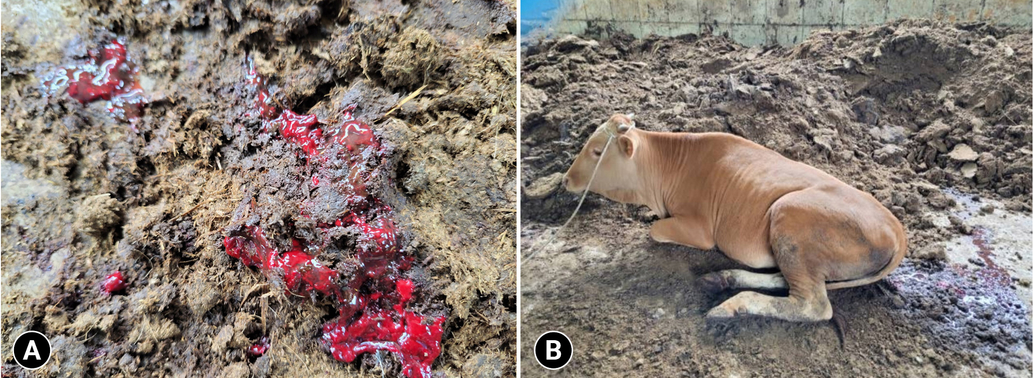
Clinical symptoms of the steer (case 2). Diarrhea with blood and mucus (A) and depression and astasia (B) were observed.
After the necropsy, tissue samples (abomasum, ileum, cecum, colon, rectum, and kidney) were collected. Blood samples from 23 cattle in the same herd were further collected using K2 EDTA spray-dried anticoagulated blood collection tubes Becton Dickinson, NJ, USA. The laboratory, the blood samples were centrifuged at 3,000 g for 20 minutes to separate the plasma and used it for BVDV screening. Total RNA was extracted from plasma and tissue samples using the RNAiso Plus Reagent (Takara Bio, Japan) and RNeasy Mini Kit (Qiagen, Germany), respectively. RT-PCR was performed using the 5ʹ-UTR, with a predicted size of 288 bp. The amplicons were subjected to direct sequencing (Macrogen, ROK). All sequences obtained were analyzed using Chromas and constructed using MEGA X. The nucleotide sequence generated in this study was deposited in the GenBank database with the accession number OQ275005.
The remaining tissues were fixed in 10% buffered formalin, routinely processed and embedded in paraffin for hematoxylin and eosin staining for routine histological examination. All evaluations were performed by trained investigators (Animal and Plant Quarantine Agency; APQA, ROK). All tissues used for histological analyses were also used for immunohistochemistry (IHC) to detect the BVDV Ag (APQA).
Antibody for BVDV was measured using a commercially available IDEXX BVDV Ab Test kit (IDEXX Laboratories, Switzerland) per the manufacturer’s instructions. The presence or absence of BVDV Ab in the sample is determined by the sample/positive (S/P) ratio for each sample. The S/P ratio for individual plasma samples was calculated using the manufacturer’s formula. Samples were considered negative if they had S/P ratios of < 20 and positive if they had S/P ratios of ≥ 30.
All samples collected from the steer were found to be positive for BVDV. Sequencing analyses revealed that this animal was confirmed to be infected with BVDV2a (Supplementary Fig. 1). The sequence obtained from this steer showed 97.2% to 97.9% homology with those reported in the ROK. In other cattle being raised in the same herd, the BVDV Ag was not detected, whereas all animals that were examined tested positive for the BVDV Ab.
Gross examination revealed that the abomasum had erosions and extensive hemorrhage in the mucosa (Fig. 2A), with mucosal ulceration, inflammation, and hemorrhage in the pylorus (Fig. 2B). The ileum was covered with brown and bloody fluids (Fig. 2C); however, other gross findings, such as erosion and petechiae, were not found. Extensive petechiae and blood clots were observed in the mucosa of the cecum and colon (Fig. 2D and E). The colon also displayed extensive hemorrhage.

Gross lesions observed during the necropsy. (A) Erosion (arrow) and extensive hemorrhage in the abomasum mucosa. (B) Ulceration (arrow) and inflammation of the pylorus. (C) Bloody and brown fluids (arrow) in the ileum. (D) Extensive petechia in the colon. (E) Diffused hemorrhages and clotted blood (arrows) of the colonic mucosa.
Histopathologic lesions confirmed ulcerative abomasitis in the pylorus. Although there was no focal gastric structure, inflammatory exudates remained in the pylorus (Fig. 3A). Hemorrhagic necrotizing enteritis was noted in the ileum. Epithelial cells of the intestinal mucosa were destroyed and macrophages containing hemosiderin infiltrated the ileum (Fig. 3B). In the colon and rectum, erosive enteritis was observed. Moreover, necrosis of scattered cells deep in the epithelium, crypt dilatation, and cell debris were observed in the crypt cells of these tissues (Fig. 3C and D). IHC analysis revealed that the BVDV Ag was detected in all tissues, except for the kidney. In particular, the colon and rectum exhibited higher rates of BVDV Ag positivity than the abomasum and small intestine (Fig. 3E-H).

Histological (A-D) and immunohistochemistry (E-H) images of the digestive tract. (A) Lack of focal gastric structures and the remaining inflammatory exudate. (B) Damaged epithelial cells and infiltrated macrophages (arrow) in the ileum. (C) Erosive enteritis in the colon. (D) Necrosis within the epithelium and crypt dilatation in crypt cells of the rectum. (E-H) Bovine viral diarrhea virus Ag (arrows) was widely distributed in all tissues, especially in the abomasum (E) and ileum (F). Tissues were also stained with primary anti-BVDV antibody (DMAB28412; Creative Diagnostics, USA) and examined by light microscopy (E-H). Scale bars: A, B, and E, 100 ㎛; C, D, and F-H, 200 ㎛.
The present case describes severe hemorrhagic syndrome caused by BVDV2a. The abovementioned clinical and pathological findings, such as severe hemorrhagic diarrhea, extensive mucosal hemorrhage, erosion, and ulceration shown in this steer were similar to those in previously reported MD cases [11,12]. However, dermatitis, which is frequently present in MD, was not noted in this steer, whereas severe erosions and ulcerations in the upper digestive tract shown in this case were typical of MD. The occurrence of MD requires the presence of a PI animal congenitally infected with BVDV. Currently we are unsure whether this steer was PI because viremia was not tested. Considering that BVDV was not detected in serum samples from other cattle in the same herd, this steer is likely not PI; rather, it was acutely infected with BVDV. Unfortunately, because viral isolation was not performed, it cannot be confimred whether this steer was infected with the cp or the ncp virus. However, sequencing analysis confirmed that there were no changes in the viral genome, such as gene duplication, deletion, and insertion in the sequence detected [6]. Moreover, IHC results revealed marked positive staining for BVDV Ag in the large intestine, whereas it was not detected in the kidney. This provides the main key that differentiates PI from an acute/transient infection. Thus, our findings suggest that this steer was acutely infected with ncp BVDV2.
Ncp BVDV2 strains have also been associated with acute hemorrhagic enteritis [13], which is consistent with our findings. BVDV2 is generally considered more virulent because it is frequently associated with diseases and death [14]. Notably, unlike the animal that died earlier, this steer exhibited astasia (Fig. 1). We are unsure whether the virus might affect the brain. The immunosuppressive effect of BVDV may play an important role in exacerbating the pathogenic effect of other infectious agents in cattle [15]. This herd was not vaccinated against BVDV. The infection route of the virus within the herd is uncertain. Per our observation, new animals had been introduced because this farmhouse has a system of breeding cattle for a short period and selling them. This might act as the key factor for viral transmission.
This report describes a case of hemorrhagic syndrome caused by ncp BVDV2a infection. The diagnosis was based on the pathological features and results of IHC and RT-PCR analyses. In the outbreak reported, the clinical and pathological presentations were found to be acute. The clinical signs were severe hemorrhagic diarrhea, depression, and astasia. Gross lesions were mainly mucosal erosion, ulceration, and petechiae. Viral antigens were more intensely detected in the large intestine. The economic impact of the outbreak was substantial owing to the acute infection and sudden death of two steers over a short period.
Notes
The authors declare no conflict of interest.
Acknowledgements
This research was funded by the Korea Institute of Planning and Evaluation for Technology in Food, Agriculture, and Forestry (IPET) (Grant No. 122017-02-1-HD020).
Supplementary Materials
Supplementary data are available at https://doi.org/10.14405/kjvr.20230005.
Supplementary Fig. 1.A phylogenetic tree based on the 5'-untranslated region was generated using MEGAX and the maximum likelihood method; the numbers over branches indicate the bootstrap values (1,000 replicates) as percentages, supporting each phylogenetic branch. The sequence identified in this study is shown in boldface. BVDV, bovine viral diarrhea virus.