토끼 고환에서 발생한 과립세포종 2예
Testicular granular cell tumor of two rabbits: case reports
Article information

Trans Abstract
Granular cell tumor was described in the testis of two rabbits. Testis from each rabbit was surgically removed and submitted for histopathological diagnosis. Both testes were about 2.0 cm in diameter, firm, and tan. Microscopically, testicular mass consisted of compact sheets of round to polygonal and occasional spindle-shaped cells. The neoplastic cells contain a large amount of eosinophilic granular material in the cytoplasm. The cytoplasmic eosinophilic granules were positive for periodic acid Schiff stain. Immunohistochemically, the neoplastic cells were immunoreactive to Melan-A and vimentin. Based on these results, the testicular mass was diagnosed as a granular cell tumor.
과립세포종(granular cell tumor)은 동물에서 비교적 드물게 발생하는 종양으로 조직학적으로 세포질에 호산성의 과립을 함유하고 있는 것이 특징이다[1,2]. 과립세포종은 대부분 양성이며, 구강 특히 혀, 피부, 뇌, 심장, 암컷 및 수컷 생식기 등 다양한 부위에서 발생이 보고된 바 있다[1,3–7]. 말의 경우는 특징적으로 거의 대부분 폐에서 발생이 보고되었으며, 랫드의 경우는 뇌막에서 발생하는 종양의 82% 정도가 과립세포종이라고 한다[3,5,7]. 과립세포종의 정확한 기원에 대해서는 아직까지도 정확하게 규명이 되지 않았다. 과거에는 미분화된 근섬유에서 유래하는 것으로 알려졌으나, 최근에 면역염색성을 근거로 Schwan cell에서 유래하는 것으로 간주되고 있다[8,9].
토끼 고환 종양에 대한 후향적 연구는 2건이 보고되어 있다[2,8]. 그 중 1건은 고환 종양은 토끼에서 발생하는 종양 중 1.93%를 차지하였고, 1.93% 중 간질세포종이 81.4%를 차지한다고 보고하였다[8]. 다른 후향적 연구에서는 토끼 고환 종양이 매우 드문 종양이나 가장 많이 발생하는 고환 종양은 과립세포종(고환 종양 중 63% 차지)이라고 보고하였다[2].
토끼에서 고환 종양에 대한 보고 사례는 드문 것으로 알려져 있으며, 발생률, 품종 특이성, 잠복 고환과의 관련성, 치료 및 회복, 종양의 분류 등도 확실히 정립되어 있지 않다[8]. 지금까지 보고된 토끼의 고환 종양은 간질세포종(Leydig cell tumor), 정상피종, 기형종, 성선모세포종, 세르토리세포종(Sertoli cell tumor), 과립세포종이 있으며, 이중에서 간질세포종과 세르토리세포종이 주를 이루고 특히 간질세포종이 가장 많이 진단되었다[8].
토끼에서의 간질세포종과 과립세포종은 hematoxylin and eosin (H&E) 염색상에서 형태학적으로 매우 유사하기 때문에 정확한 감별진단이 필요하다. 감별진단을 위한 방법으로 periodic acid Schiff (PAS) 염색과 면역조직화학적 염색, 전자현미경적 방법이 있다. 이 사례들은 토끼에서 매우 드물게 발생하는 과립세포종을 PAS 염색법과 면역조직화학적 염색법을 실시하여 진단하였기에 보고하고자 한다.
각각 2살 및 3살된 라이언 헤드(lionhead), 더치(Dutch)종인 수컷 토끼로, 고환은 대략적으로 2주에서 1개월가량의 경과를 보이며 점차적으로 종대되었다. 신체검사 시에 음낭 피부의 병변은 관찰되지 않았고, 편측의 고환만이 커져 있었으며 음낭 내에서 자유롭게 움직였다. 유선의 발달, 주변 림프절의 종대와 같은 소견은 관찰되지 않았다. 그러나 2마리 중 1마리는 경도의 음낭 수종이 관찰되었다. 음낭 수종을 보인 개체는 혈액학적 검사에서 림프구 감소 소견을 보였으며, 혈액화학검사에서 alkaline phosphatase (ALP)와 glucose의 증가를 보였다. 반면에 다른 개체는 혈액학적 검사에서 특이한 변화는 관찰되지 않았지만, 혈액화학검사에서 ALP, glucose, 나트륨의 증가를 보였다. 의뢰된 2마리 고환의 크기는 대략 2 × 2 cm 정도였으며, 단면은 거의 대부분 유백색조의 종괴로 대체되어 있어 정상적인 고환의 모습은 소실되어 있었다(Fig. 1). 고환은 각각 외과적으로 절제한 다음 병리조직학적 진단을 실시하였다.
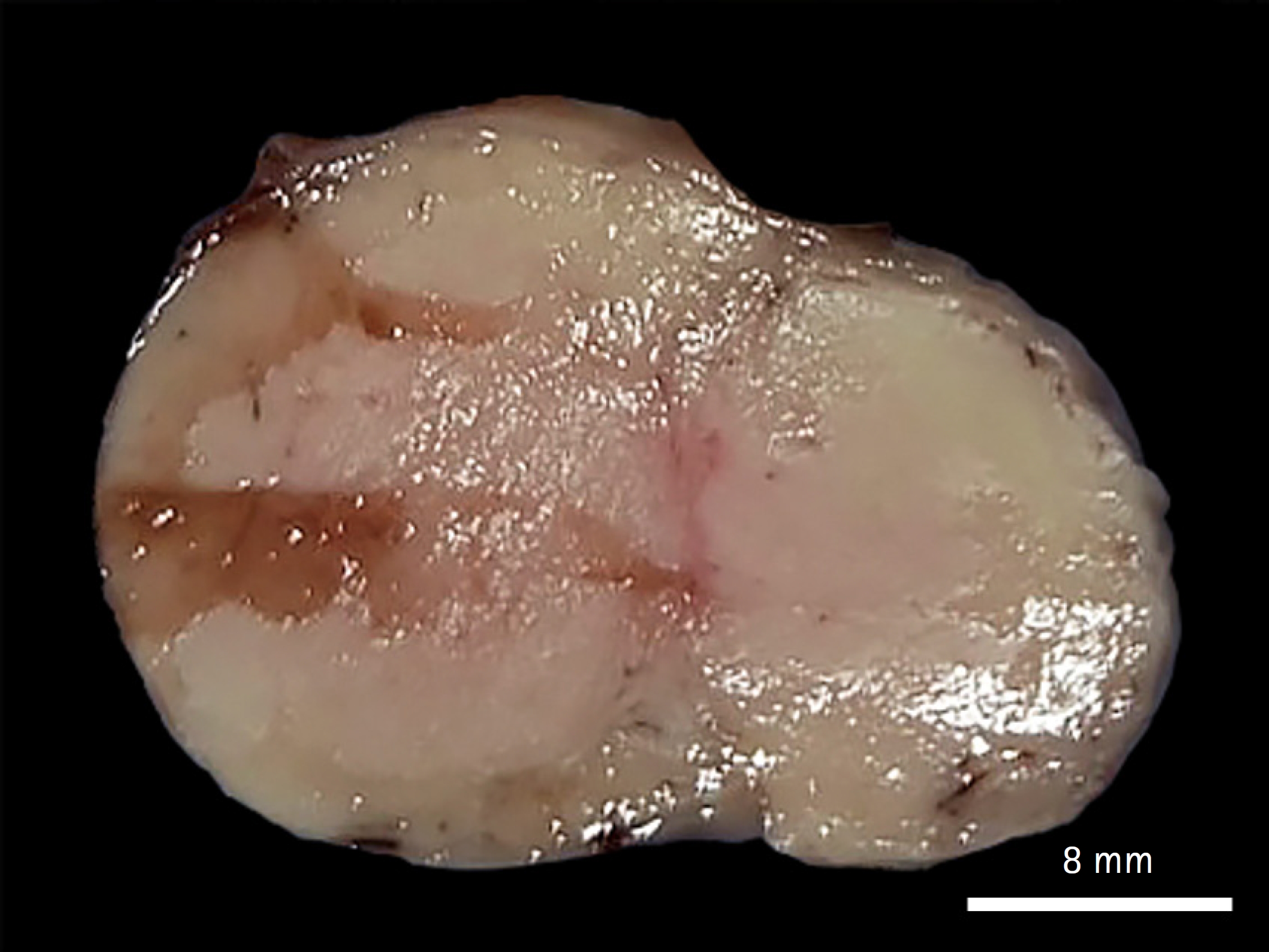
Most of the testicle in rabbits was replaced with a whitish mass, and the normal appearance of the testicle was lost (scale bar = 8 mm).
절제한 종괴는 병리조직학적 진단을 위해 10% 중성 포르말린에 충분히 고정시키고 통상적 조직처리 과정을 거친 다음 파라핀에 포매한 후 4 μm 두께로 절편을 만들어 H&E 염색을 실시하였다. 연속 절편 조직에서 과립세포종을 증명하기 위해 PAS 염색을 실시하였고, 종양세포의 기원을 밝히기 위해 Melan-A (dilution 1:50, mouse, monoclonal, No. M7196, A103; Dako, USA)와 vimentin (dilution 1:200, mouse, monoclonal, No. M0725, V9; Dako)에 대하여 면역조직화학 염색을 실시하였다. 면역조직화학 염색용 조직 절편을 citrate buffer (pH 6.0)에 넣어 heat-induced antigen retrieval을 실시하였고, 내인성 과산화효소를 제거하기 위해 3% 과산화수소 용액에 처리하였다. 각 항체를 4℃에서 24시간 동안 반응시킨 후에 host species-matched horseradish peroxidase (HRP)-결합 이차항체를 상온에서 2시간 동안 반응하였다. 발색제로 3,3-diaminobenzidine solution를 사용하였고 Mayer’s hematoxylin으로 대조 염색을 실시하였다. 양성대조군으로는 흑색종 조직(Melan-A 항체)과 정상 장 조직(vimentin 항체)을 사용하였다.
병리조직학적으로 의뢰된 두 고환 조직들은 거의 유사한 병리조직학적 소견을 보였다. 고환 종괴에서는 원형에서 다각형 형태의 종양세포들이 주를 이루었고, 드물게 방추형의 종양세포들이 촘촘한 시트상(sheet)으로 증식하고 있었다(Fig. 2A). 종양세포의 세포 경계는 뚜렷하였으며, 풍부한 호산성의 과립성 세포질을 가지고 있었다(Fig. 2B). 종양세포의 핵은 원형에서 타원형이었으며, 유사분열상은 거의 관찰되지 않았다. 종양세포 세포질에서 관찰되었던 호산성의 과립들은 거의 대부분 PAS 염색에 양성을 보였다(Fig. 3A), Vimentin과 Melan-A 항체에 대한 면역조직화학적 염색 결과, 양성대조군과 마찬가지로 두 항체 모두에서 종양세포 세포질에서 갈색으로 강한 양성반응을 나타내었다(Fig. 3B and C).
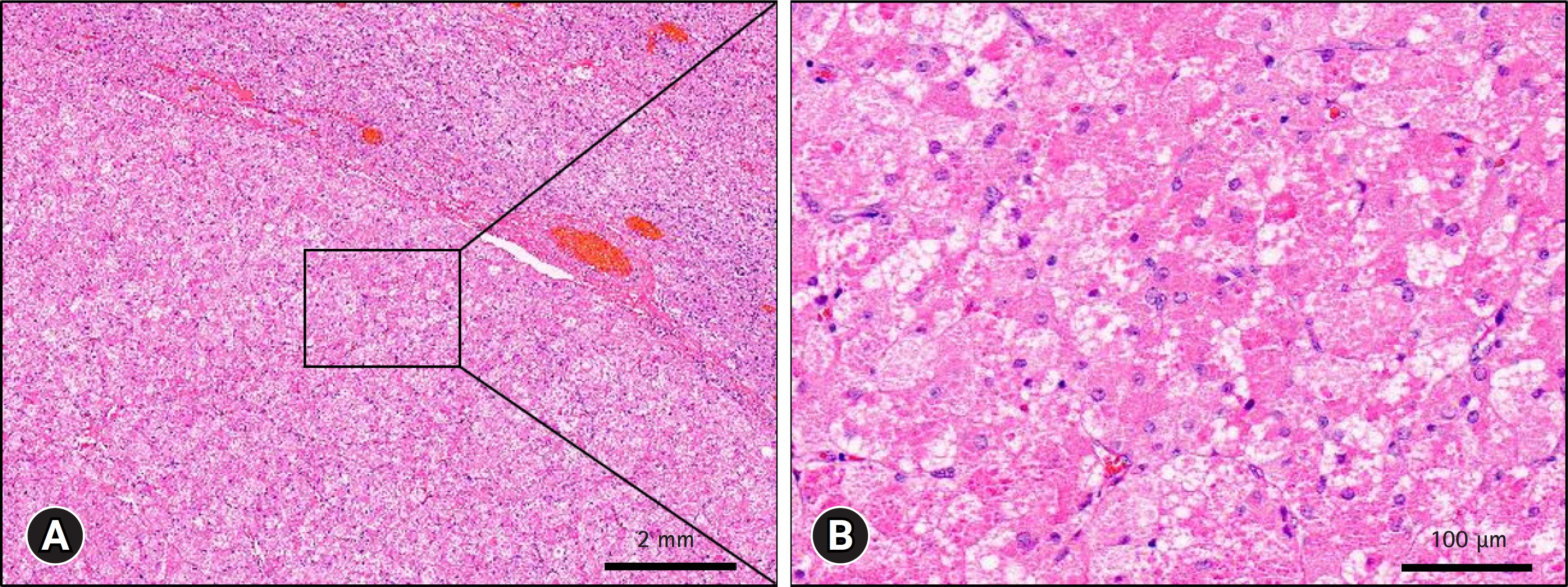
Testicular granular cell tumor in rabbits. Representative photomicrograph of a hematoxylin and eosin stain. The majority of the testes were replaced by neoplastic mass, obliterating the normal histologic architecture of the testis (A). The cell boundaries of the neoplastic cells were distinct and had abundant eosinophilic granular cytoplasm (B). Scale bar: (A) 2 mm, (B) 100 μm.
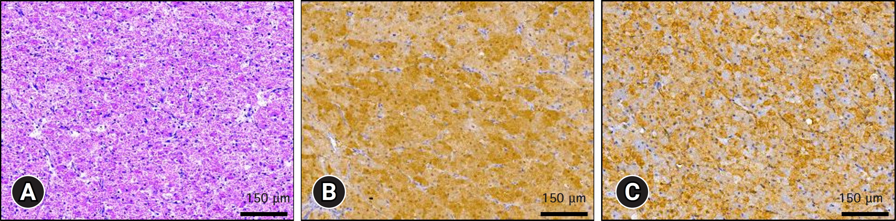
Testicular granular cell tumor in rabbits. Representative photomicrograph of a periodic acid Schiff (PAS) stain (A) and immunohistochemical stain for Melan-A (B) and vimentin (C). The cytoplasmic eosinophilic granules were positive for PAS stain (A). Neoplastic cells showed positive immunoreactivity for both Melan-A (B) and vimentin (C). (A–C) Scale bars = 150 μm.
과립세포종은 형태학적으로 간질세포종과 유사하기 때문에 H&E 염색을 통한 일반적인 병리조직학적 검사뿐만 아니라 PAS 염색과 면역조직화학적 염색이 병행하여야 한다고 알려져 있다[2,8]. PAS 염색은 두 종양의 감별진단에 매우 유용한 방법으로 보고되었다[2,8,10]. PAS 염색은 조직 내에 있는 글리코겐, 당단백질 또는 중성 당을 포함하는 점액물질을 검출하는 방법으로 사람에서 과립세포종, 폐와 신장의 투명세포종, 림프육종, 난소의 브렌너종양, 정상피종, 중피종의 진단을 위해 사용되었다[11]. 과립세포종에서 대부분의 종양세포는 PAS 양성을 나타내었으나, 간질세포종을 포함하는 다른 성삭기질 종양의 종양세포는 음성이었다[10]. 이번 사례에서도 PAS 염색에서 대부분의 종양세포가 양성반응을 보였다. 또한, 개, 고양이, 말, 앵무새와 같은 동물의 과립세포종에서도 PAS 염색 결과는 유사한 경향을 보였다[10]. Webb 등[8]은 기존에 병리조직학적으로 간질세포종으로 진단되었던 53건 중 29건을 PAS 염색을 통해 과립세포종으로 재분류하였다.
대부분이 과립세포종은 신경외배엽에서 유래한 것으로 알려져 있어[9], 이번 사례에서는 정확한 진단을 위해 PAS 염색 이외에 신경외배엽 관련 항체(Melan-A)와 vimentin을 이용하여 면역화학염색을 실시하였다. Melan-A 항체와 vimentin 항체에 대해서는 모든 종양세포가 양성을 나타내었으나, neuron-specific enolase 항체에 대해서는 10% 정도의 종양세포만이 양성을 나타냈다[1]. 그러나 Melan-A 항체에 대한 양성반응은 간질세포종과 같은 스테로이드 호르몬을 생산하는 성삭기질 종양에서도 나타나는 것으로 알려져 있어[12], 진단에 주의가 필요하다. 앞서 열거한 2가지 항체 외에도 periaxin 항체와 S100 항체를 이용한 면역화학염색이 과립세포종의 진단에 사용되기도 하였다[2]. 그러나 Melan A와 vimentin 항체의 반응성보다는 낮았다.
이번 사례의 2건의 토끼 고환 종양은 호산성의 과립성 세포질을 가진 원형에서 다각형 형태의 종양세포가 주를 이루어 병리조직학적으로 전형적인 과립세포종의 특징을 보였다. 또한 간질세포종과 구별하기 위해 실시한 PAS 염색에서 대부분의 종양세포는 양성반응을 나타내었고, Melan A와 vimentin 항체에 대한 면역조직화학 염색에서 모든 종양세포는 양성반응을 나타내어, 이들 결과들을 바탕으로 토끼에서 발생한 고환 과립세포종으로 진단하여 보고하고자 한다.
Notes
The authors declare no conflict of interest.
Acknowledgements
본 논문은 2023년도 교육부의 재원으로 한국연구재단의 지원을 받아 수행된 지자체-대학 협력기반 지역혁신 사업의 결과이며, 이에 감사드립니다(2021RIS-001 [1345370811]).