

Introduction
Modern nanotechnologies have helped change many of the medical rules used to prevent, diagnose, and treat diseases. This technology offers new ways for agents to cross the cell membrane and differentiate between target and non-target cells in the human body. Gold nanoparticles (AuNPs) have been studied widely for the treatment of diseases, particularly cancer. AuNPs (5 to 100 nm in size) have been applied since the Middle Ages, which led to the name potable gold [1]. Medically, AuNPs are used in radiomedicine and have a high X-ray absorption coefficient and local surface plasmon resonance [2]. In addition, AuNPs exhibit electronic and optical properties that allow controllable interactions with organic molecules, including electron donor groups [3]. Gold has been used to treat neurological disorders, such as depression, epilepsy, migraine, and gland disorders, such as menopause and sexual dysfunction. On the other hand, the real effect of gold was not understood physiologically for a long time because of the apparent contradiction that gold is a rare metal with virtually no toxicity [4,5]. While gold toxicity has recently been demonstrated, unlike most pharmaceuticals, it is not generally related predictably to the limits it reaches within the body tissues [5].
Cystic hydatidosis is a zoonotic disease of humans. Infection of the intermediate hosts results from ingesting Echinococcus granulosus eggs in contaminated water and food, which develop into the hydatid cysts, mainly inside the liver or other body tissues. Dogs, wolves, and foxes are considered the definitive hosts of E. granulomas [6,7]. This disease is characterized by the absence of pathological symptoms in the initial stages of infection. With time, the cysts grow in size inside the organ, leading to pressure on the affected organ and its neighboring organs [6,8]. Patients typically exhibit an inflammatory reaction. In advanced cases, the hydatid cyst may obstruct the function of the affected part. On the other hand, various immune reactions, such as urticaria, edema, and anaphylactic shock, occur when the cyst ruptures and the hydatid fluid is released, possibly resulting in death [9-11].
The complete failure of surgical therapy was one of the reasons that prompted researchers to use many other methods to treat it, such as chemotherapy, immunotherapy, radiotherapy, and treatment using medicinal plants. Recently, some nanomaterials have been studied to determine their effect on the vitality of protoscolices outside the living body. This study evaluated the scolicidal effect of AuNPs against protoscolices of hydatid cysts.
Materials and Methods
Hydatid cysts and protoscolices preparation
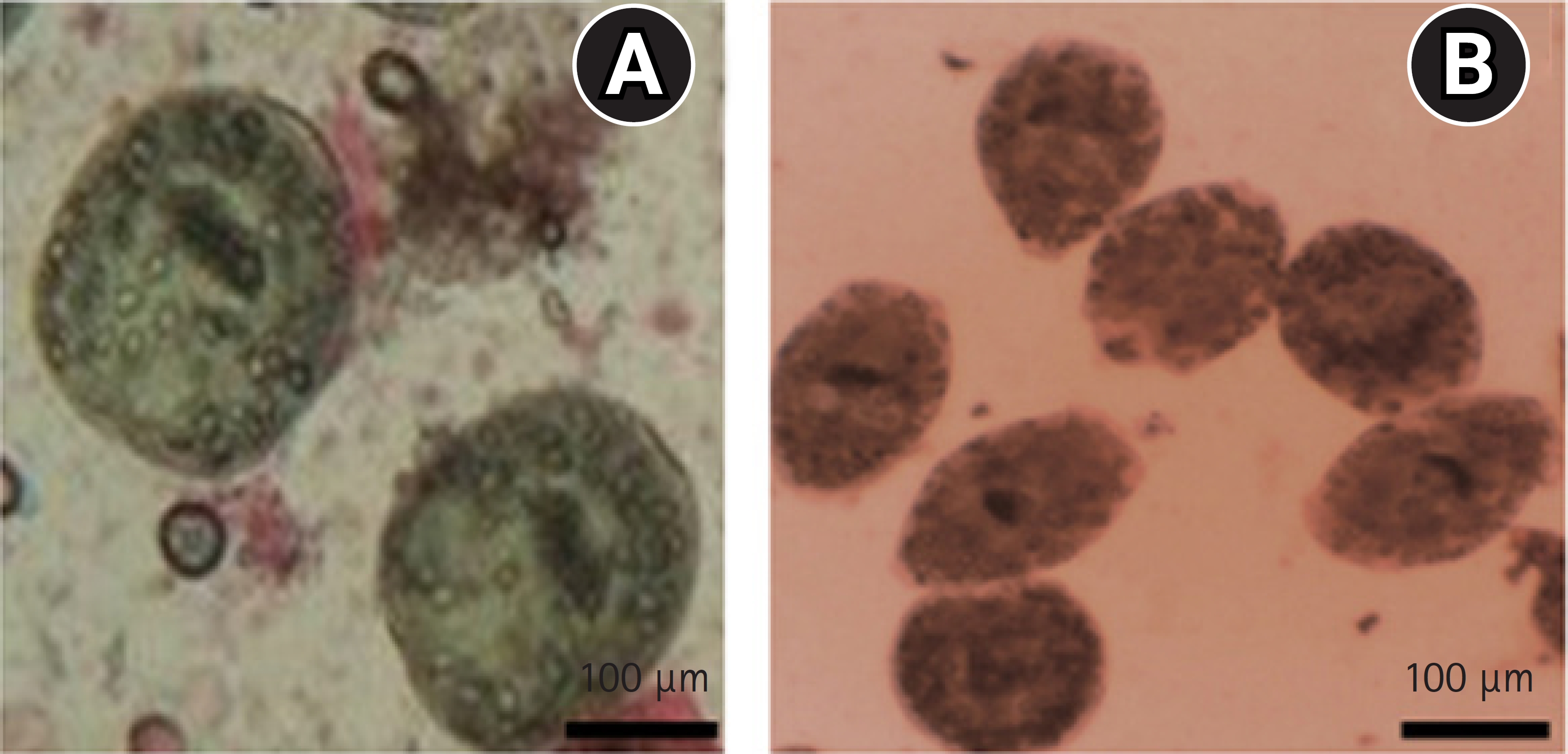
Hydatid cysts were collected from naturally infected sheep from the Diwaniyah abattoir in Diwaniyah Province, Iraq. The cysts were washed from the outside with tap water to remove blood and suspended matter accumulated during slaughter and placed inside a clean container. They were then transferred to the parasitology laboratory at the Department of Biology, Faculty of Education, University of AL-Qadisiyah. The protoscolices were aspirated under sterile situations and saved in sterile tubes. The vitality of the protoscolices was assessed by taking 1.5 mL of the protoscolices suspension and staining with 1% eosin. The solution was shaken well, and a drop was taken from it and examined under a microscope. The live protoscolices appeared green compared to the dead protoscolices, whose membranes had been penetrated by the dye and dyed red [12].
Preparation of AuNPs
AuNPs were obtained as ready-made oxide as a gold powder with a purity of 99.9% and a particle size of 30 to 60 nm (Sky Spring Nanoparticles Inc.). The AuNPs were prepared from a stock solution (2 g of AuNPs per liter of distilled water) and sterilized in an autoclave. The solution was mixed using an ultrasonic homogenizer for 15 minutes, after which 0.2, 0.4, 0.8, and 1 mg/mL colloids were prepared and kcept in the refrigerator until use.
Effectiveness of concentrations of AuNPs 0.2, 0.4, 0.8, and 1.0 mg/mL on the vitality of protoscolices in vitro
In this step, 1.5 mL of the suspension of the protoscolices was transferred to 20 tubes. The tubes were then treated with 0.2, 0.4, 0.8, or 1 mg/mL of gold nanoparticles for 15, 30, 45, or 60 minutes. The arithmetic average of the protoscolices vitality was calculated under the above conditions, as evidenced by the permeability of eosin dye. The protoscolices were examined by optical microscopy. The live and dead protoscolices were counted based on the dyeing of protoscolices with the eosin dye [13].
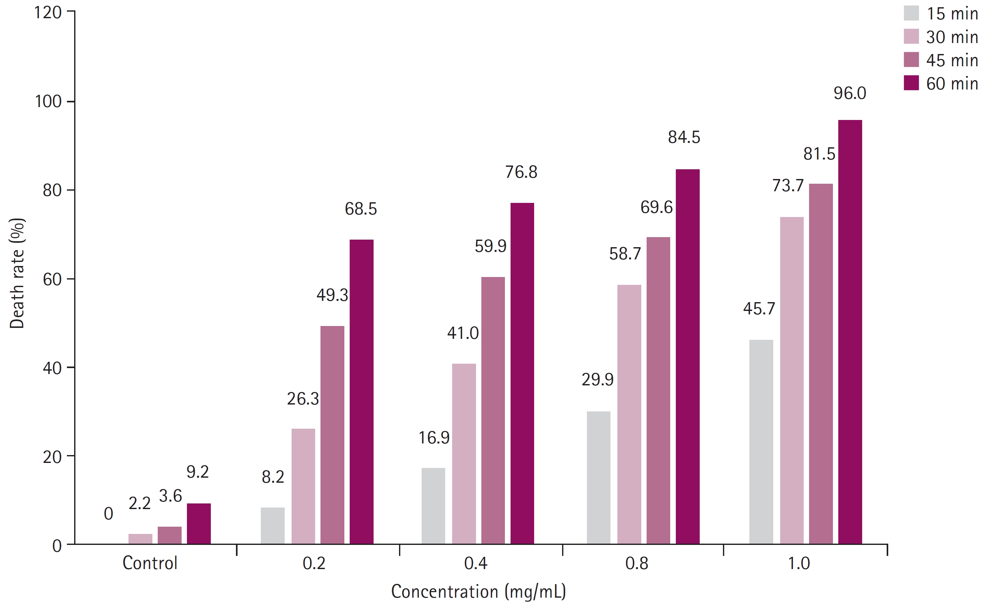
Results
The percentage of protoscolices destruction increased as the concentration and duration of exposure to AuNPs increased (Table 1 and Fig. 1). The percentage destruction was 68.5%, 76.8%, 84.5%, and 96.0% at 0.2, 0.4, 0.8, and 1.0 mg/mL, respectively, after a 60 minutes treatment, representing the highest percentage of protoscolices mortality compared to the protoscolices mortality treated with the same concentrations for shorter periods. The lowest mortality was observed in the group treated for 15 minutes: 8.2%, 16.9%, 29.9%, and 45.7% at 0.2, 0.4, 0.8, and 1.0 mg/mL, respectively. The mortality rate increased with time. The death rate after 30 minutes was 26.3%, 41.0%, 58.7%, and 73.7% at 0.2, 0.4, 0.8, and 1.0 mg/mL, respectively. After 45 minutes of treatment, the death rate of the protoscolices was 49.3%, 59.9%, 69.6%, and 81.5% at 0.2, 0.4, 0.8, and 1.0 mg/mL, respectively. In contrast, the control group showed a 0%, 2.2%, 3.6%, and 9.2% death rate at 15, 30, 45, and 60 minutes, respectively (p<0.05). Overall, the AuNPs caused significant morphological changes to the protoscolices, as shown in Fig. 2.
Discussion
Hydatid disease is a widespread disease with an estimated prevalence of 5% to 10% [14,15]. According to the World Health Organization, Iraq has a high endemicity of hydatid cyst disease. Clinical studies have shown that treatment using drugs does not always kill the protoscolices [16], and surgical methods are not devoid of risks, such as the rupture of hydatid cysts and spillage of their contents [17]. Therefore, research is continuing to discover effective and safe methods. AuNPs were used in this study as a scolicidal agent because of the presence of previous studies that demonstrated their effectiveness against various parasites, such as protozoa comprising [18] Toxoplasma gondii, Trypanosoma spp., Leishmania spp. [19], and Cryptosporidium spp.; helminths comprising trematodes (Schistosoma spp.) [20] and cestodes (Raillietina spp.) and vectors containing mosquitoes [21].
Eosin stain penetration into the primary primates was used to verify the vitality of protoscolices because eosin penetration is a permeable process related to the nature of the permeability of the membrane. In contrast, live protoscolices retain their natural green color [22,23]. Four concentrations of AuNPs were used (0.2, 0.4, 0.8, and 1.0 mg/mL) with 0.9% physiological saline used as the control group. The results indicated that the death rate of the protoscolices increased directly as the concentration and duration of exposure to the AuNPs increased.
The death rates of the protoscolices exposed 0.2, 0.4, 0.8, and 1.0 mg/mL for 60 minutes were highest at 68.5%, 76.8%, 84.5%, and 96%, respectively. By contrast, the lowest percentage of protoscolices death after 15 minutes of treatment was 8.2%, 16.9%, 29.9%, and 45.7%, respectively. This result is consistent with ├ćolak et al. [24], who reported that AuNPs have a scolicidal effect and can treat cysts associated with the bile ducts. In addition, another study [25], which used four concentrations of AuNPs, reported that AuNPs have promising results as a scolicidal agent for cystic hydatid cysts.
The 1 mg/mL concentration was the optimum because it led to a 96% death rate after 60 minutes owing to the inhibitory effect of gold nanoparticles, which may have affected the physiology of the protoscolices through its effect on enzymes or stopping the cell metabolism cycles that occur within the protoscolices. The AuNPs lead to paralysis and then the death of worms. The authors attributed this to changes in the enzymatic activity of the worms [26] in addition to its effect on a group of protozoa [27-29] and the morphological changes in the protoscolices. The morphological distortions of the protoscolices exposed to gold nanoparticles were attributed to the loss of the plasma membrane function or defects in the equilibrium of ions on both sides of the membrane plasma.
 PDF Links
 PubReader
 ePub Link
 Full text via DOI
 Download Citation
 Print
Print


