Ultrasonographic diagnosis of calcifying tendinopathy of the biceps brachii in a Doberman Pinscher dog: a case report
Article information

Abstract
A 10-year-old, spayed female, Doberman Pinscher dog presented with right forelimb lameness. On physical examination, painful reaction and crepitation were present at the right shoulder. Radiographic evaluation of the shoulder revealed nothing remarkable. On the ultrasonography of the right shoulder joint, 2 small echogenic masses accompanied by reverberation artifacts were observed in the right biceps tendon near its origin at the supraglenoid tuberosity. Based on these findings, we suspected biceps calcifying tendinopathy. Clinical signs resolved intermittently after administration of nonsteroidal anti-inflammatory drug. This study described the ultrasonographic findings of calcifying biceps brachii tendinopathy which is an unusual finding in dogs.
The biceps brachii muscle attaches proximally at the supraglenoid tubercle by means of the long tendon of origin (the origin of biceps brachii tendon). This crosses the shoulder joint in a sharp curve to reach the cranial surface of the humerus through the intertubercular groove. In the elbow joint region, the tendon of insertion (insertion of biceps brachii tendon) splits into 2 parts [1]. Bicipital calcifying tendinopathy is defined as dystrophic calcification of the tendon of origin of the biceps brachii muscle [2].
Degenerative or traumatic conditions in dogs involving the shoulder joint may cause forelimb lameness associated with the biceps tendon [3]. Although tendon trauma or rupture may cause calcifying tendinopathy, the exact pathogenesis is still unclear [4]. Calcifying biceps brachii tendinopathy is rare in dogs [5]. Large and medium-sized dogs are mainly affected. Labrador retrievers and Rottweilers are particularly overrepresented [5]. Most dogs with calcifying tendinopathy are chronically or intermittently lame and at times are asymptomatic [6,7].
A diagnosis of calcifying tendinopathy can be based on a combination of physical examination, radiography, ultrasonography, computed tomography, or magnetic resonance imaging (MRI) [5]. A radiographic examination can reveal calcification overlying the craniomedial aspect at the approximate position of the humerus [4]. Ultrasonography is sensitive and accurate techniques for tendon evaluation [8]. The most common ultrasonographic finding was hyperechoic area accompanied by distal acoustic shadowing [3,8]. A heterogeneous tendon pattern and hypoechoic area around calcification may be observed in the calcifying tendinopathy [8]. There was evidence that the presence of a hypoechoic area surrounding the calcification was related to clinical signs of pain, suggesting an active inflammatory process [8]. Ultrasonography can provide detailed information about the size, shape, location, and presence of an inflammatory change surrounding the calcifying lesion [8]. For an accurate diagnosis of shoulder joint conditions, histopathological examination with excisional biopsy is essential [9]. This report describes the ultrasonographic features of calcifying tendinopathy in the biceps tendon, an infrequent condition of shoulder joint diseases in dogs.
A 10-year-old spayed female Doberman Pinscher dog with right forelimb lameness was referred to the Gyeongsang National University veterinary medical teaching hospital. The dog presented having chronic and intermittent right forelimb lameness over the previous 3 years. Abduction and extension of the right shoulder elicited a mild crepitation and painful reaction during physical examination. There were no remarkable findings on serum chemistry values or complete blood counts.
On survey shoulder radiographs, no specific findings were found in either shoulder (Fig. 1). On ultrasonography (Arietta 70; Hitach Aloka Medical, Japan), the patient was positioned in left lateral recumbency, with the right forelimb uppermost and the shoulder slightly flexed and externally rotated and a 12 MHz linear probe used to examine the tendons in transverse and longitudinal planes. Scapulohumeral joint ultrasonography found a well-defined linear-shaped bright echogenic mass accompanied by reverberation artifacts and another small echogenic mass in the right biceps tendon near its origin at the supraglenoid tuberosity. These were considered to be biceps tendon calcification. However, small gas could not be excluded. In addition, we identified an irregularly shaped, hyperechoic mass accompanied by distal acoustic shadowing between the biceps tendon and the supraglenoid tuberosity. That was consistent with the osteoarthritic supraglenoid tuberosity. We also saw a small amount of anechoic fluid beneath the tendon (Fig. 2). Based on the ultrasonographic examination, the tentative diagnosis was biceps calcifying tendinopathy. However, the possibility of biceps tenosynovitis or tendinitis could not be ruled out because a histopathologic examination was not performed in this dog.
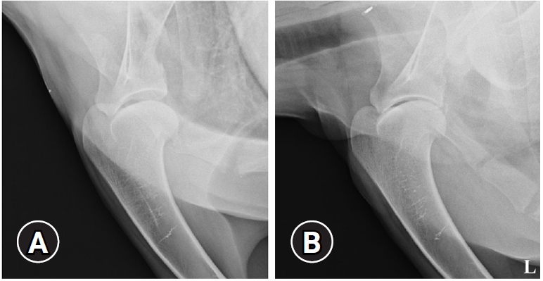
Mediolateral radiographic view of the (A) right and (B) left scapulohumeral joint of the patient. No remarkable findings were confirmed on radiography. L, left.
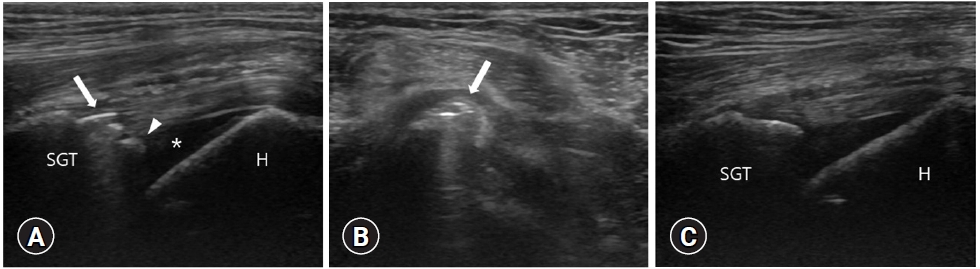
Ultrasonographic images of the patient’s scapulohumeral joint. (A) Sagittal image of right scapulohumeral joint. Linear bright mass accompanied by reverberation artifact in the biceps tendon (arrow) was identified. Irregularly shaped, hyperechoic masses with distal acoustic shadowing (arrowhead) were confirmed on the outside of the tendon. A small amount of anechoic fluid (asterisk) was seen beneath the tendon near its origin at the supraglenoid tuberosity. (B) Transverse image of right scapulohumeral joint. Linear bright masses (arrow) was also seen in this view. (C) Sagittal image of left scapulohumeral joint. A seen small amount of anechoic fluid was also seen beneath the biceps tendon. SGT, supraglenoid tubercle; H, humerus.
The dog received nonsteroidal anti-inflammatory drug (carprofen 4.4 mg/kg single in day, per oral) for 14 days. Additionally, a behavioral restriction was implemented for 6 weeks. Clinical signs of right shoulder problems such as pain and lameness gradually improved. Ten months later, the dog expired, and this was presumed to be because of an underlying dilated cardiomyopathy.
Calcifying tendinopathy is known to occur idiopathically in horses and humans, and is rare in dogs, mainly found in large and medium-sized dogs especially in Rottweilers and Retrievers [6,10]. In this case, the dog was Doberman Pinscher, which is large breed of dog.
One study shows that the prevalence of calcifying tendinopathy in the scapulohumeral joint in dogs is 7%, and such dogs present as being either chronically or intermittently lame, or asymptomatic [6]. Bicipital tenosynovitis and tendon calcification in dogs are not always associated but may be connected from the standpoint of pathological processes [9]. In humans, although the etiology and pathogenesis of calcifying tendinopathy are not clear, it may be that tendon trauma or rupture can reduce the blood supply and cause hypoxia [11].
In dogs, radiographic imaging in the contralateral limb is useful for direct comparison, and the cranioproximal-craniodistal view of the shoulder helps to verify brachii tendon calcification [12]. Calcifications overlying the craniomedial aspect of the approximate position of the humerus can been seen on radiography [4]. In this case, only the lateral and caudocranial views were available, and no specific radiographic findings were identified.
Ultrasound is known to be more accurate than radiography in tendon assessment [13]. In dogs, it is well known that when the shoulder joint is flexed, it is the easiest to assess the biceps brachii tendon, which is easily palpable within the bicipital groove of the greater tubercle of the shoulder joint. For perpendicular orientation of the transducer to the tendon fibers, it is necessary to apply slight pressure with the transducer [3]. In this case, we maintained flexion of the shoulder joint and had no difficulty in evaluating the biceps brachii tendon.
In dogs, the biceps tendon can be readily identified on ultrasonography as it displays dense linear, and hyperechoic fiber patterns. The biceps tendon is homogenous and hyperechoic from its origin to its musculotendinous junction. On transverse imaging, this tendon typically varies from a circular to a flat shape as it goes from its insertion distally [3]. Furthermore, dystrophic mineralization can be recognized as discrete, focal, reflective interfaces with distal acoustic shadowing in the tendon [3]. In this case, 2 echogenic masses were confirmed in right biceps tendon, and they were considered to be a calcification of the biceps tendon.
A small accumulation of anechoic fluid is visible below the origin of the supraglenoid tubercle tendon, but this may also be seen in biceps tenosynovitis in dogs [3,9]. In this case, on ultrasonography, a small amount of anechoic fluid was also identified beneath the tendon near its origin at the supraglenoid tuberosity. Although a histopathological examination could not be performed, it was considered normal because it had also been confirmed on the contralateral limb.
The limitation of this case is that histopathologic examination was not performed. However, there were no remarkable findings to indicate inflammation on radiography or ultrasonography. Thus, we assumed that tendinopathy was more likely than either tendinitis or tenosynovitis. Arthroscopy of the scapulohumeral joint may be useful for diagnosing by direct visualization of the origin of the biceps brachii tendon and proximal intertubercular groove [14]. MRI is thought to be the best diagnostic imaging tool for evaluating both the intra- and extraarticular structures in dogs [15]. However, neither arthroscopy nor MRI was done in this case.
The case described the ultrasonographic findings of calcifying tendinopathy of the biceps brachii tendon. Although it is a rare disease in dogs, calcifying tendinopathy of the biceps brachii tendon should be considered in dogs with shoulder lameness and ultrasonography can be useful for diagnosis of calcifying tendinopathy of the biceps brachii tendon.
Notes
The authors declare no conflict of interest.