

Introduction
The usage of herbal plants as medication for the treatment of different diseases have been harvested by humankind for at least 60,000 years (aspirin from willow tree bark and morphine from opium). It is due to the fact that nature has bestowed healing properties in plants and even allopathic drugs are synthetic analogs of various active compounds present in plants [1,2].
Lespedeza cuneata (LC) or Chinese bush clover is common in Asian countries and widely used as traditional medicine. Bioactive compounds isolated from LC include pinitol, phenol, tannin, ╬▓-sitosterol, quercetin, kaempferol, vitexin, and orientin [3,4]. LC is effective in the treatment of prostatic hyperplasia [5], early atherosclerosis [6], and as skin whitening agent [7].
Coal fly ash (CFA) is a significant contributor to air pollution, resulting from industrial activities and vehicular emissions in urban areas. CFA comprises of liquid and solid particles of varying sizes and origins, including coal, combustion particles, and asbestos [8]. Nanoparticles from CFA can persist in the atmosphere for extended periods and easily disperse in the air. Chronic exposure to CFA can result in the accumulation of sediment in the lungs, leading to severe adverse effects such as inflammation in the alveolar cells [9,10].
Inflammation is the first defense response mechanism by the body against pathogen invasion. Inflammatory reactions are essential for the maintenance of homeostasis in the body. However, dysregulated inflammation can amplify reactive oxygen species (ROS) production and release inflammatory mediators [11,12]. MH-S cells, which are alveolar macrophages derived from the mouse lung, produce a substantial proportion of nitric oxide (NO) via inducible NO synthase (iNOS) upon stimulation with LPS or foreign invaders [13]. Moderate amounts of NO can be neutralized by intercellular vascular regulation, the immune system, and signal transmission. In contrast, excessive amounts of NO can cause septic shock, cerebral infarction, diabetes, and degenerative neurological diseases [14,15].
MH-S cells produce different cytokines, together with tumor necrosis factor (TNF)-╬▒, interleukin (IL)-1╬▓, and IL-6 and critically regulate pro-inflammatory cascade under the induction of an endotoxin or CFA [16]. TNF-╬▒ affects the vascular endothelium and endothelial leukocytes [17]. IL-1 ╬▓ is an initiator of cyclooxygenase-2 (COX-2), and IL-6 is a regulator of immune reactivity [18]. Overproduction of these cytokines contributes to several inflammatory disorders. Therefore, it may be possible to prevent diseases by inhibiting the production of inflammatory mediators.
Even though the anti-inflammatory activity of LC has been reported in RAW 264.7 murine macrophage cell line and paw edema in rats [19], research data on CFA-induced inflammation is not reported with LC. Therefore, we geared to check the anti-oxidative and anti-inflammatory effects of LC on MH-S cell line induced with CFA. The anti-inflammatory activity was checked by NO levels and pro-inflammatory mediators and cytokines expression via polymerase chain reaction (PCR), while the antioxidant activity was measured by 1,1-diphenyl-2-picrylhydrazyl (DPPH) and 2,2'-azino-bis(3-ethylbenzothiazoline-6-sulfonic acid) (ABTS) assays. Cell viability was determined by methyl thiazolyl tetrazolium (MTT) assay.
Materials and Methods
Chemicals and reagents
Roswell Park Memorial Institute medium, fetal bovine serum (FBS), penicillin-streptomycin, and DulbeccoŌĆÖs phosphate buffered saline were obtained from Welgene Co. (Korea). CFA, 3-(4,5-dimethylthiazol-2-yl)-2,5-diphenyltetrazolium bromide (MTT) and dimethyl sulfoxide (DMSO) were procured from Sigma-Aldrich (USA). Oligo-dT (Bioneer oligo synthesis), COX-2, iNOS, TNF-╬▒, IL-1╬▓, and IL-6 primers were obtained from Bioneer. TRIzol reagent was purchased from Invitrogen (USA). All other chemicals and reagents used were of laboratory and cell-culture grade.
Sample preparation of LC
LC was extracted for 2 hours with distilled water at 100┬░C, 50%, 70%, and 100% EtOH at 80┬░C in a 1:20 ratio (plant: solvent w/v). After extraction, these samples were filtered using Whatman filter papers. The samples were then evaporated via rotary evaporator and placed at -70┬░C overnight to freeze. The samples were then freeze-dried for 3 to 4 days at -55┬░C to obtain powdered yield. The powdered samples were weighed. DMSO was used as solvent for ethanol extracts in particular concentrations for the evaluation of the samples.
Gas chromatography-mass spectrophotometry analysis
To determine the single compounds in the extracted powdered sample, GC-MS analysis was performed using an Agilent 7890A GC instrument (Agilent Technologies, USA). The instrument was equipped with a 30ŌĆēmŌĆē├ŚŌĆē0.25ŌĆēmm (i.d. DB-5MS) chromatography column and an Agilent 5975C mass selective detector. The extract was injected at a temperature of 250┬░C. The source and transfer line temperatures were set at 230┬░C and 280┬░C, respectively. The column temperature was initially set at 70┬░C for 1 minute and then increased at a rate of 5┬░C/min to a final temperature of 300┬░C, which was maintained for 30 minutes. Mass spectrometry data was obtained in scan and electron ionization modes to analyze the LC compounds. The data for single compounds obtained is shown in Table 1.
DPPH assay
To analyze anti-oxidative effects, ABTS and DPPH assays were performed as described previously [20]. Briefly, 20 ┬ĄL of LC at different concentrations of 7.8 to 1,000 ╬╝g/mL was reacted with DPPH reagent, side by side with ethanol (control). Incubated for 30 minutes at 37┬░C, absorbance was checked at 517 nm. Radical scavenging activity was calculated using the formula, while ascorbic acid (AA) serving as the positive control:
DPPH scavenging activityŌĆē(%) = [1 ŌłÆ (A1 ŌłÆ A2)/(A3 ŌłÆ A4)] ├Ś 100
where A1 is the absorbance of DPPH and the sample, A2 is the absorbance of 100% ethanol and the sample, A3 is the absorbance of DPPH and the solvent used for sample dilution (DMSO/DDW), and A4 is the absorbance of 100% ethanol and the solvent used for sample dilution (DMSO/DDW).
ABTS assay
Free radicals were created for the ABTS assay by allowing 14 mM ABTS solution (5 mL) to react with 4.9 mM potassium persulfate (K2S2O8) solution (5 mL), which was then incubated at room temperature (22┬░CŌłÆ26┬░C) for more than 12 hours. An absorbance of 0.700 ┬▒ 0.020 at 734 nm was kept the target absorbance before starting assay. LC 50 ╬╝L at different concentrations 7.5 to 1,000 ╬╝g/mL was reacted to 100 ╬╝L of the final ABTS solution at various concentrations keeping Trolox as positive control. Radical scavenging activity can be calculated as:
ABTS scavenging activityŌĆē(%)ŌĆē= [1 ŌłÆ (A1 ŌłÆ A2)/(A0)] ├Ś 100
where A1 is the absorbance of the ABTS working solution and sample, A2 is the absorbance of the sample without ABTS working solution, and A0 is the absorbance of the ABTS working solution only.
Cell culture
Murine alveolar macrophage cell line MH-S, originating from the American Type culture collection, was cultured in DMEM supplemented with 10% FBS (WelGene Co.) and 100 IU/mL penicillin and 100 ╬╝g/mL streptomycin sulphate (Lonza, USA). The incubating conditions were humidified 5% CO2 at 37Ōäā.
Nitric oxide assay
NO levels were measured using Griess reaction assay as described previously [21]. Briefly, MH-S cells were seeded in 6-well plates for 24 hours, then stimulated with CFA (2.5 ╬╝g/mL) for 18 hours, with or without LC 70% EtOH at the indicated concentrations of 50 to 400 ╬╝g/mL. After 18 hours cell culture supernatants were reacted with Griess reagent for 5 minutes at room temperature (22┬░CŌłÆ25┬░C) and the absorbance of each well was then measured at 540ŌĆēnm using a microplate reader (VersaMax Microplate Reader, USA).
Cell viability (MTT) assay
MTT reagent was added to the cells at a concentration of 0.1 mg/mL in order to measure cell viability. After the NO analysis, cells were exposed to the MTT reagent for 3 hours. The resulting violet crystals were then dissolved in DMSO, and the absorbance at 560 nm was measured using VersaMax Microplate Reader.
Reverse-transcription PCR and real-time PCR
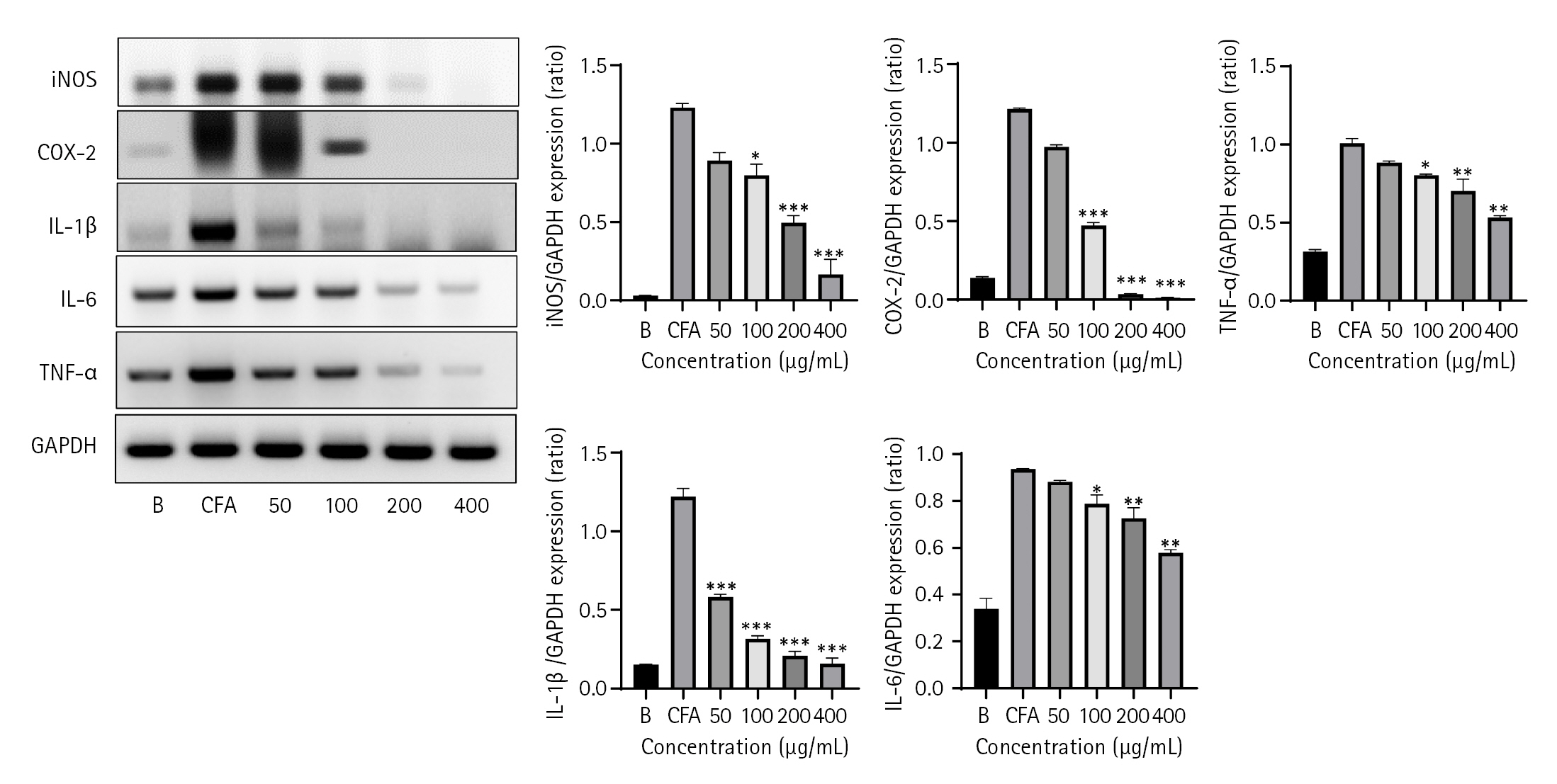
Following a 30-minute pretreatment with or without LC 70% EtOH at the given concentrations of 50 to 400 ╬╝g/mL MH-S cells were stimulated with CFA (2.5 ╬╝g/mL) for 18 hours. Total RNA was extracted using the TRIZOL reagent after 18 hours in accordance with the manufacturerŌĆÖs instructions. Reverse transcription was carried out using a reverse transcriptase premix (Bioneer), followed by annealing with Oligo-dt for 10 minutes at 70┬░C, cooling for 5 minutes on ice, and incubating for 90 minutes at 42.5┬░C using a thermal cycler (Biometra GmbH, Germany). To activate the reverse transcriptase, the processes were paused at 95┬░C for 5 minutes. The cDNA aliquots obtained from this process were utilized for reverse-transcription (RT)-PCR, and the PCR results were then electrophoresed on a 1% agarose gel. Ethidium bromide was used to stain the gel, and Eagle Eye image analysis software was used to identify bands (Stratagene, USA). The band intensities were normalized to that of the corresponding GAPDH bands as housekeeping gene. Primers sequence used for RT-PCR and real-time PCR analysis are mentioned in Table 2.
Statistical analysis
Data are represented as means ┬▒ standard error of the mean. DunnettŌĆÖs test and one-way ANOVA were used to statistically analyze the data. Differences were tested using statistical analysis software (ver. 9.4; SAS Institute, USA), and statistical significance was set at p < 0.001.
Results
LC exhibits potent free radicals scavenging activity with DPPH and ABTS assay
LC exhibited strong radical scavenging activity, which decreased in a dose-dependent manner (Fig. 1A). At a sample concentration of 1,000 ╬╝g/mL, the radical scavenging activity of LC was similar to that of AA in the DPPH assay. In contrast, using Trolox as a positive control, the sample exhibited higher radical scavenging activity with the ABTS assay than with the DPPH assay at similar concentrations (Fig. 1B). Our results indicate that LC exerts more effective results with the ABTS assay, demonstrating that LC has strong anti-oxidative activity because of the presence of phenolic and flavonoid components.
LC reduces NO production without cytotoxicity in MH-S cells
NO is an inflammatory product produced by cells invaded by foreign pathogens. In our experiment, MH-S cells were treated with various doses of LC and, after 30 minutes of stimulation with CFA 2.5 ╬╝g/mL, as an inducer of inflammation, NO was produced in MH-S cells, as visualized using Griess reagent at 540 nm. Fig. 2A depicts NO production and dose-dependent reduction by LC from 50 to 400 ╬╝g/mL. NO concentration decreased significantly from 100 ┬Ąg/mL of the sample and was lowest at 400 ╬╝g/mL.
Cell viability was assessed using the MTT assay to evaluate whether the dosage used was cytotoxic. As depicted in Fig. 2B, LC did not exert any cytotoxicity, and its anti-inflammatory activity did not affect cell viability.
Effects of LC on inhibition of inflammatory cytokines using RT-PCR and Real-time PCR
To assess the inhibition of the inflammatory cytokine profile, we performed RT-PCR (Fig. 3). After measuring NO production, RNA was extracted from MH-S cells, cDNA was produced, amplified through PCR, and gene expression was assessed using gel electrophoresis. As shown in Fig. 3, mRNA expression of inflammatory mediators and cytokines, such as iNOS, COX-2, IL-1╬▓, IL-6, and TNF-╬▒, decreased significantly with increasing concentrations of the sample. These results indicate that LC inhibits inflammation by reducing the production of inflammatory mediators and cytokines.
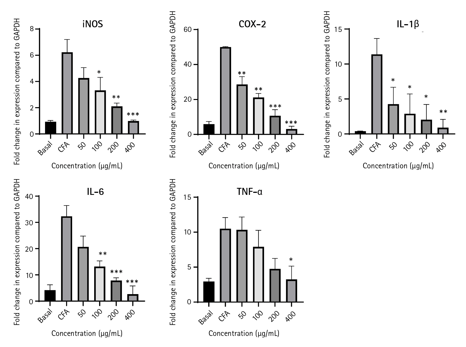
We also performed real-time PCR to determine the fold change in the gene expression of each inflammatory cytokine. LC significantly suppressed inflammatory cytokines and mediators i.e. iNOS, COX-2, IL-1╬▓, IL-6, and TNF-╬▒ (Fig. 4).
Discussion
Almost all physiological abnormalities have been treated traditionally using natural remedies. Owing to the scientific progress, we can now examine the molecular processes that underlie the effects of the substances present in such natural products (herbs, shrubs, roots, leaves, and flowers). In Korean traditional medicine, LC has been used to treat acute mastitis [22], leukorrhea, asthma, stomach ache, and diarrhea [19]. It has also been reported for it beneficial effects in treatment of testicular dysgenesis syndrome by increasing sperm count and muscle mass in andropause rats [23]. Furthermore, LC and its single compounds are reported for anti-oxidation [24], anti-diabetic and insulin secretion enhancer [25], cytoprotection from ROS [26], tyrosinase inhibitor (skin whitening agent) [27] and as antibacterial agent [28].
Unstable free radicals can be stabilized in the body by the redox-functionalized proton ions supplied in DPPH and ABTS radical-scavenging tests, which are essential for this process. This is typically accomplished by exploiting the fact that antioxidants cause unstable violet DPPH and ABTS free radicals to change into stable yellow DPPH free radicals by absorbing a hydrogen ion [29]. DPPH was used to examine the antioxidant activity of compounds and extracts as its absorption at 517 nm decreases with the presence of antiradical substances [30]. We also evaluated the formation of ABTS cations through the ABTS-potassium persulfate reaction as a marker of the antiradical chemicals present in the samples [31]. A previous study on plums reported a strong link between antioxidant activity determined by the ABTS assay and total phenolic content but a poor correlation between antioxidant activity and total flavonoid content [32]. Conversely, ABTS radicals function via electron transfer, whereas DPPH radicals are engaged in transferring hydrogen atoms. Fig. 1 demonstrates that our plant extract contained more phenolic components and produced more effective results with the ABTS assay, demonstrating that LC has a strong anti-oxidative activity because of the presence of phenolic and flavonoid components. This result is supported by literature that phenols and flavonoids are strong antioxidants in nature [33].
Cells produce NO as a defense mechanism against foreign compounds and pathogens invasion. Typically, NO is a gaseous moiety that aids cells in fending off external invaders; however, NO is continuously created during the persistent invasion, thereby harming the adjacent cells [34]. Therefore, the primary action mechanism for most anti-inflammatory medications from allopathic or herbal sources is timely management of NO production. In the present study, we demonstrated that LC effectively reduced NO levels in MH-S cells activated in vitro with CFA (Fig. 2). Severe infection and sepsis are a matter of concern to patients and physicians. Viral or bacterial infections are frequent causes of infection. Pro-inflammatory chemicals, cytokines, and anti-inflammatory substances are generated in response to infection. The body can eliminate invasive pathogens and is shielded from damage caused by inflammation because of the delicate balance between pro- and anti-inflammatory chemicals [35].
Typically, NO synthesis is considered as a protective mechanism against endotoxic shocks, such as that against CFA. iNOS and COX-2 are 2 pro-inflammatory mediators produced when CFA binds to Toll-like receptor 4, activating downstream inflammatory signaling pathways. These pro-inflammatory mediators activate the cascade of pro-inflammatory cytokines, including IL-1╬▓, IL-6, and TNF-╬▒. Notably, these 3 cytokines may be harmful to their cells if their release is not effectively inhibited [36]. This notion is further confirmed by our results (Figs. 3, 4). Under the LC treatment mRNA expression of pro-inflammatory mediators and cytokines, including iNOS, COX-2, IL-1╬▓, IL6, and TNF-╬▒, decreased significantly with increasing concentrations, as assessed through RT-PCR and real-time PCR, to alleviate CFA-induced inflammation.
CFA, a by-product of burning coal, is a synthetic source of fine particle matter (PM). The largest coal-burning nation in the world, China, has seen a 5.5-year decline in life expectancy partly owing to respiratory illnesses caused by exposure to high coal-combustion PM levels [37]. The purpose of providing this information here is to corrleate CFA with respiratory system anomalies. CFA in conjuction with other air pollutants encourage the permeability of lungs epithelial cells to viral receptors. This in turn leads to the suppression of host cells defensive mechansims towards antigen processing and T-cell functions. Our previous studies have shown that natural herbal extracts, such as those of Duchesnea indica, Salvia plebeia, and Aster scaber inhibit CFA-induced inflammatory activity in in vitro and in vivo asthma models, respectively [16]. Similarly, our current results indicated that LC is a potent herbal plant extract that inhibits CFA-induced inflammation in murine alveolar cell lines by inhibiting the expression of pro-inflammatory mediators and cytokines. Therefore, it may have potential as supplement in the treatment of respiratory diseases (e.g., severe acute respiratory syndrome coronavirus 2).
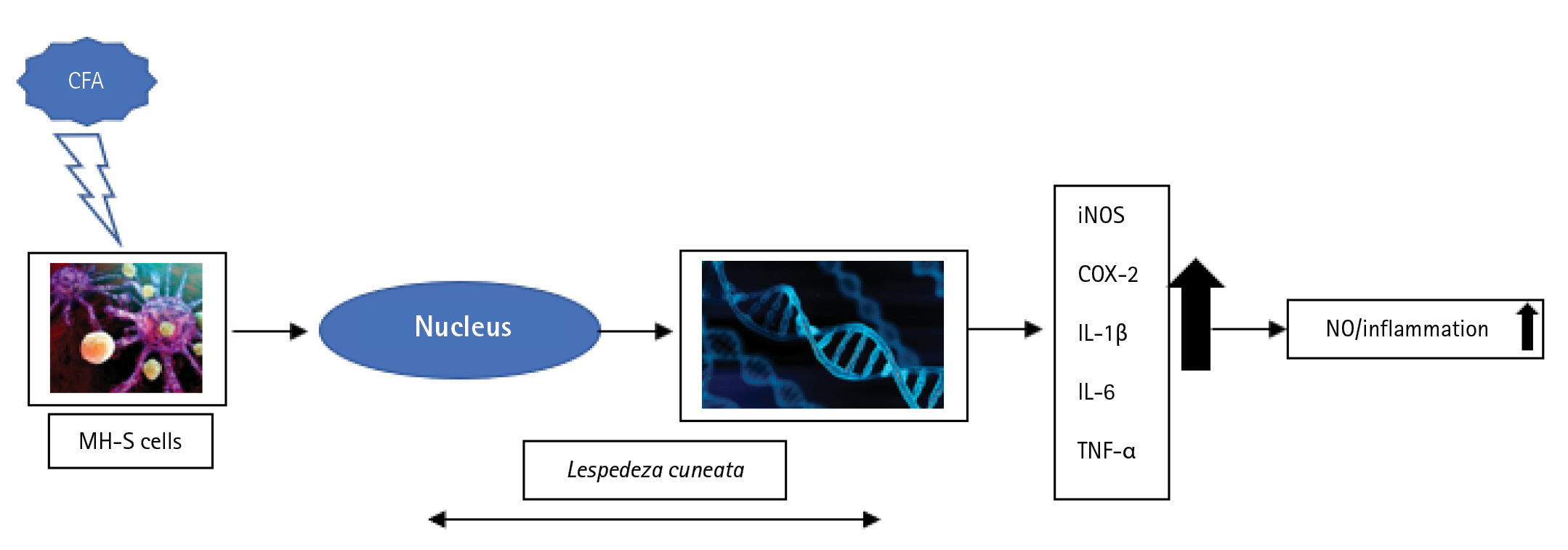
Natural products have been extensively used for years in the past. However, scientists have evaluated natural products containing equivalently potent substances compared with that of synthetic drugs without side effects. LC is commonly distributed in Asian countries and reportedly has therapeutic effects. Our results demonstrated that LC has strong anti-oxidative effects via inhibiting free radicals produced by the DPPH and ABTS assays. Collectively, our results demonstrate that LC exerts potent anti-inflammatory effects by inhibiting NO production via different pro-inflammatory mediators and cytokines, i.e., iNOS, COX-2, IL-1╬▓, IL-6, and TNF-╬▒ as shown in Fig. 5.
In summary, LC can be a potent nutraceutical drug candidate for commercial use and investigated further for its therapeutic anti-oxidative and anti-inflammatory effects.


 PDF Links
 PubReader
 ePub Link
 Full text via DOI
 Download Citation
 Print
Print


