Korean J Vet Res > Volume 62(4); 2022 > Article
Seung, Kim, Kim, Kim, Jang, Kim, Her, Yi, Lee, Jang, and Lee: Serological responses and protection levels in chickens administered with Newcastle disease vaccines

Abstract

Vaccination against Newcastle disease (ND) is the most effective means of controlling the disease, and these vaccines are commercialized only after their safety and effectiveness have been verified through tests that comply with Korean Standards of National Lot Release for Veterinary Biologics. This study investigated whether a relatively convenient and safe serological test can be used in place of the challenge test using highly virulent ND virus. Hemagglutination inhibition (HI) assay and enzyme-linked immunosorbent assay (ELISA) were considered positive of log2 2 or more and cutoff value of 200 or more, respectively, in both live and inactivated vaccines. However, when the antibody levels of the live and inactivated vaccines induced using the Ulster 2C, KBNP-C4152R2L, and K148/08 strains were compared, the antibody titers for inactivated vaccines were significantly higher than those for live vaccines in both the HI assay and ELISA. A strong positive correlation was observed between HI and ELISA antibody titers. The live vaccines corresponded to a survival rates of ≥ 80% and the inactivated vaccines corresponded to 100% survival rates. This study confirmed that standard efficacy tests can serve as serological tests, and can replace the challenge test and that the vaccine approval process can be improved.

Introduction

Newcastle disease (ND) is one of the most critical infectious diseases in domestic and wild birds [1]. Generally, ND primarily causes acute respiratory disease; however, it can also lead to depression, nervous manifestations, and diarrhea [2]. ND virus (NDV) or Avian paramyxoviruses 1 (APMV-1) was recently classified to Family Paramyxoviridae, Genus Avian Orthoavulavirus and common NDV is recently known as Avian Orthoavulavirus-1 (AOAV-1) [3]. Although only a single serotype of NDV has been found to be exist as determined using neutralizing tests and cross-protective analysis [4,5], the virus can be categorized into 2 major classes based on phylogenetic analysis of the fusion (F) gene sequence, i.e., Classes I and II [3]. Moreover, Kapczynski et al. [6] reported that NDV is constantly evolving and that it can be classified into more than 21 phylogenetically distinct genotypes as per epidemiological evidence. Class I NDVs are genetically less diverse, are present in wild waterfowl, and have low virulence; in contrast, Class II NDVs are more diverse, frequently isolated from poultry with occasional spillovers into wild birds, and have a wide range of virulence. Moreover, Class II NDV can be subdivided into 21 genotypes [1,4,7].
Generally, virulent NDV can be defined as NDV with an intracerebral pathogenicity index of ≥ 0.7 in 1-day-old chicks or multiple basic amino acids at the cleavage site of the virus F protein, according to the World Organisation for Animal Health (OIE) [8]. NDV isolates can be classified into 3 major pathological types based on virulence: velogenic (high virulence), mesogenic (moderate virulence), and lentogenic (low virulence) [9]. Velogenic NDV can be subdivided into viscerotropic velogenic NDV (vvNDV), which causes severe visceral and intestinal hemorrhage, and neurotropic velogenic NDV, which presents with severe neurologic clinical signs, depending on the clinical signs in chicken behavior [10]. ND is one of the damaging diseases among chickens, and it has a significant global economic impact worldwide [11]. Moreover, there are no reports of treatment of ND and no antiviral drugs are commercially available.
Vaccination against ND is the most effective means of controlling the disease, and this method is practiced in most countries producing commercial and backyard poultry except Ireland, Norway, Switzerland or Sweden [12-14]. In Korea, vaccination against ND has been mandatory in hatcheries and poultry farms since 2001; consequently, no ND outbreaks have occurred since 2010 [15]. However, ND vaccination is still being used for prevention of outbreaks in the future. These vaccines are commercialized only after their safety and effectiveness have been verified through tests that comply with Korean Standards of National Lot Release for Veterinary Biologics [16]. Although vaccine validation is currently performed as a challenge test in chickens using the highly virulent NDV, this study investigated whether a relatively convenient and safe serological test can be used in place of the challenge test.

Materials and Methods

Experimental animals and NDV vaccine

Overall, 11 NDV vaccines from 6 manufacturers were tested (Table 1). The age of the test animals and the dose and inoculation route of the vaccines for evaluating vaccine strains were in accordance with the Korean Standards of National Lot Release for Veterinary Biologics [16] and the manufacturers’ recommendations. Namely, 6 vaccine strains among 7 live vaccine strains were inoculated by spray route using an automatic spray device (SKmos2009; Samkwang, Korea) in each of the 25 1-day-old specific-pathogen-free (SPF) chickens (Namduck SPF, Korea). The other live vaccine strains were inoculated by orally using a 1-mL syringe in 10 3-weeks-old and 10 6-weeks-old SPF chickens, respectively. Four inactivated vaccine strains were intramuscularly injected into 25 6-weeks-old SPF chickens. All experiments were performed thrice, and the mean outcomes were considered. The vaccinated and control groups were placed in separate isolation units throughout the study. All procedures in this study were conducted in accordance with the Institutional Animal Care and Use Committee (IACUC), APQA (approval ID: 2019-452).

Serum collection

To detect NDV antibodies, blood samples from the different groups administrated with the live (n = 10/group) and inactivated (n = 10/group) NDV vaccines were collected at 2 and 3 weeks after immunization, respectively, based on Korean Standards of National Lot Release for Veterinary Biologics [16]. Serum was separated and subjected to heat-induced inactivation at 56°C for 30 min. The samples were stored at −20°C until further analysis.

Challenge experiment

The vvNDV strain Kr005 was used as the challenge virus in this study [17]. To determine the concentration of Kr005, 50% egg infectious dose (EID50) was measured using the Reed-Muench method [18]. Virus challenge involved intramuscular injection of Kr005 (1 × 10⁵ EID50/0.5 mL) at 2 and 3 weeks after the administration of live and inactivated vaccines, respectively. The challenged chickens were observed daily for clinical signs, mortality for 2 weeks.

Hemagglutination inhibition assays

Hemagglutination inhibition (HI) assay was performed using U-bottomed 96-well microtiter plates as recommended by the OIE manual of standard diagnostic tests [8]. The assay was performed using 4 hemagglutination units of the NDV LaSota antigen. The endpoint was the reciprocal of the highest serum dilution that completely inhibited hemagglutination. HI titers were also expressed as log2.

Enzyme-linked immunosorbent assay

Indirect enzyme-linked immunosorbent assay (ELISA) was conducted to detect NDV antibodies in the serum samples using a commercial ELISA kit (MEDIAN Diagnostics Inc., Korea) according to the manufacturer’s instructions. An absorbent 450-nm filter was used to calculate the sample-to-positive (S/P) ratio of each sample using an ELISA microplate reader (Molecular Devices, USA). The presence or absence of antibodies against to NDV was determined by correlating the absorbance value of the sample to the positive control mean, and positive was indicated by an S/P ratio of > 0.2. The ELISA titer (log titer) was calculated as 1.8 (log S/P) + 3.56.

Statistical analyses

GraphPad Prism ver. 9.0 (GraphPad Software Inc., USA) was used for all statistical analyses. To compare the serological response and analyze the relationship between the antibody titers and the survival rate, independent-sample t-test and Pearson two-tailed analysis were performed. A p-value of < 0.0001 was considered statistically significant.

Results

Serological response the SPF chickens to NDV vaccine

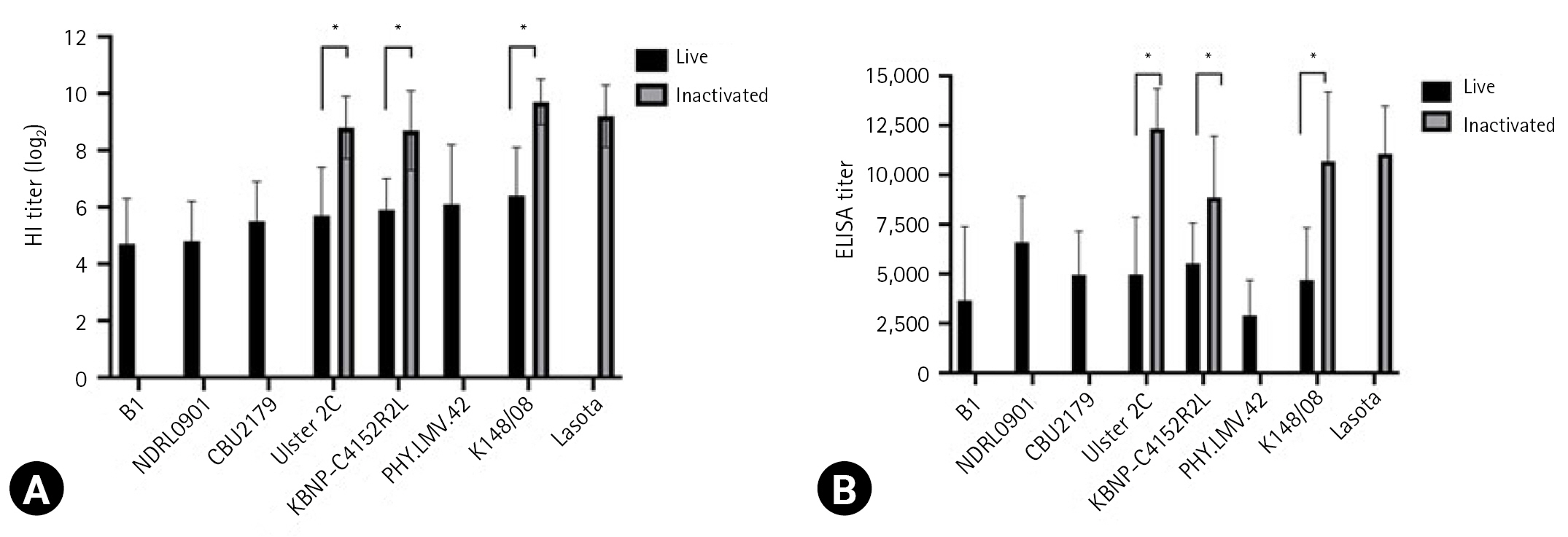
The serological response of the chickens to the vaccine strains is presented in Fig. 1. The HI assay results revealed that the mean antibody titers against the live vaccine strains ranged from log2 4.7 ± 1.6 to log2 6.4 ± 1.7, whereas those against the inactivated vaccine strains ranged from log2 8.7 ± 1.0 to log2 9.7 ± 0.8. For both live and inactivated vaccines, antibody titers were considered positive of log2 2 or more. ELISA revealed that the mean antibody titers against the live and inactivated vaccine strains ranged from 3,655 ± 3,741 to 6,601 ± 2,297 and from 8,860 ± 3,101 to 12,338 ± 2,016, respectively. All values exceeded the manufacturer’s positive cutoff value of 200. However, when the antibody levels of the live and inactivated vaccines induced using the Ulster 2C, KBNP-C4152R2L, and K148/08 strains were compared, the antibody titers for inactivated vaccines were significantly higher than those for live vaccines in both the HI assay and ELISA (p < 0.0001).

Correlation between HI and ELISA titers

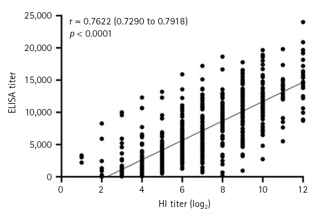
The correlation between HI and ELISA antibody titers is presented in Fig. 2. A strong positive correlation was observed between HI and ELISA antibody titers (Pearson correlation coefficient r, 0.7622; 95% confidence interval, 0.7290-0.7918; p < 0.0001).

Relationship between serological responses and protection levels

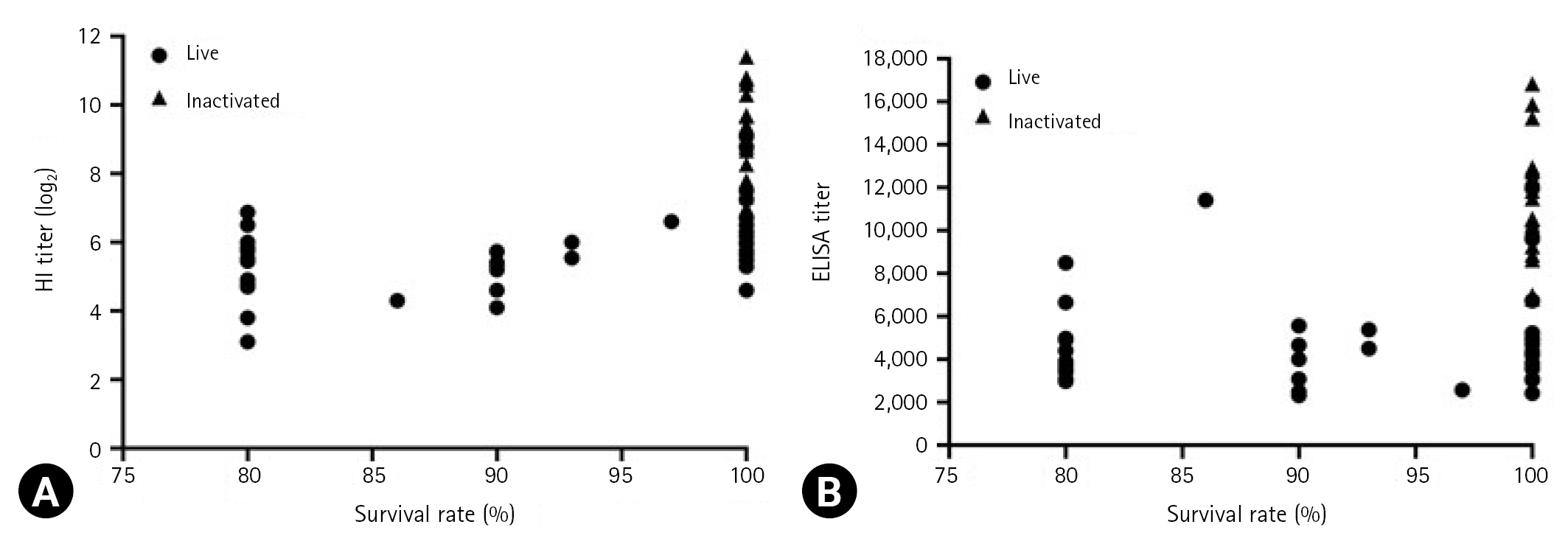
The relationship between antibody titers and survival rates is presented in Fig. 3. According to Korean Standards of National Lot Release for Veterinary Biologics, a vaccine is considered effective when the survival rates after vaccination with live and inactivated vaccines are ≥ 80 and ≥ 90%, respectively [16]. The mean HI and ELISA titers for live vaccines ranged from log2 3.1 to log2 9 and 2,316 to 11,965, respectively, corresponding to a survival rates of ≥ 80%. Further, the mean HI and ELISA titers for inactivated vaccines ranged from log2 7 to log2 11.4 and 6,808 to 16,838, respectively, both of which corresponding to 100% survival rates.

Discussion

All NDVs are of the same serotype and therefore, have similar antigenic properties; this results in a uniform immune response regardless of the vaccine strain. Both live and inactivated NDV vaccines have been widely used globally to contain the economic threat posed by ND outbreaks [6,19]. In Korea, national efficacy trials have been conducted to ensure that vaccines against ND are safe and effective in generating a protective immune response in chickens [16]. To safely manage vvNDV as a challenge virus, biosafety level 3 facilities must be developed and utilized by 2023; however, in reality, this is difficult because of problems of cost and lack of facilities in Korea [20]. Therefore, this study investigated whether the efficacy of the vaccines could be evaluated using a more convenient and safe serological test than the challenge test.
In this study, all 11 vaccines, including 7 live vaccines and 4 inactivated vaccines, achieved the positive criterion of values of log2 2 or higher in the HI assay and values exceeding the positive cutoff value of 200 in ELISA. Moreover, the HI and ELISA antibody titers generated by the inactivated vaccines were significantly higher than those generated by the live vaccines. Inactivated vaccines are generally produced as oil adjuvants to enable stimulation for a relatively long time, and they tend to increase the level of humoral antibodies in the blood after intramuscular administration [21]. Moreover, adjuvants can effectively stimulate immunity by inducing antibody production, and high antibody titers can protect against morbidity or mortality associated with NDV infection [21].
In contrast, lentogenic NDV strains are widely used as live vaccines in young birds aged 1-14 days; these vaccines block infection by inducing early immunity through cellular, local mucosal, and humoral immune responses [12]. The biggest advantage of live vaccines is that they can be administered through various routes, such as through drinking water, spray, or eye drops, and are suitable for mass distribution, resulting in extremely low costs from production to administration [6,12]. NDV comprises an envelope containing the hemagglutinin-neuraminidase (HN) protein that allows the virus to bind to host cells and the F glycoprotein that promotes the fusion of the envelope to host cells [6]. Antibodies against HN play a role in blocking virus attachment, and antibodies against the F glycoprotein inhibit virus fusion with host cell membranes, making them the primary targets for immune responses [22,23]. In this study, these antibody titers of the ND vaccine strains displayed different trends in the HI assay and ELISA, as previously described [24,25]. This is because the HI assay only detects antibodies against the HN protein, whereas the ELISA uses the whole virus as antigen, thus potentially allowing the detection of antibodies against any protein on the NDV particle [26,27]. Moreover, Thayer et al. [28] reported that ELISA could detect antibodies that did not act as protective antibodies or less analyze the role of HN in antibody induction.
However, it is impossible to unequivocally state whether this significant difference is attributable to strain characteristics. This is because although commercial vaccines contain verified vaccine strains, the composition differs. In addition, chickens vaccinated against ND produce IgM, IgY, and IgA antibodies as part of the immune response [29]. Antibodies are detected in blood 6 days after inoculation, reaching peak levels at 3 to 4 weeks resulting in high titers [30,31].
In this study, the results of the HI assay and a ready-made ELISA displayed a strong positive correlation, as previously described [32]. However, Marquardt et al. [33] reported no correlation between HI assay and ELISA. The results in this study also found that the number of positive samples was higher in the HI assay than in ELISA. In total, 0.4% of HI-negative sera samples (HI titer < log2 2) were positive on ELISA, whereas 1.7% of HI-positive sera samples were negative on ELISA (ELISA titer < 200). The results of HI assay differed from those of ELISA, and it is different from what is known that the specificity of ELISA is higher than the HI assay [32]. This discrepancy between the 2 assays may be related to the aforementioned sensitivity characteristics. Moreover, because commercial ELISA kits have different antigens for each product, differences in sensitivity and specificity between kits must also be considered. Chickens vaccinated using live and inactivated vaccines exhibited survival rates of > 80% and 100%, respectively. Thus, this study found a strong correlation between protection against challenge infection and serological titers, which is consistent with the results of previous studies [34-36]. This study confirmed that standard efficacy tests can serve as serological tests, and can replace the challenge test and that the vaccine approval process can be improved through shortening the trial periods, considering animal welfare concerns, and reducing costs.

Notes

The authors declare no conflict of interest.

Acknowledgments

This research was supported by a grant from the Animal and Plant Quarantine Agency of the Republic of Korea (no. B-1543073-2021-23-01).

Fig. 1.
Serological responses between chickens vaccinated with 2 Newcastle disease virus (NDV) groups, live and inactivated NDV vaccines. Hemagglutination inhibition (HI) titers (A) and enzyme-linked immunosorbent assay (ELISA) titers (B) were determined as the means antibody value and ± standard deviation (SD). Significant differences of antibody levels between live and inactivated vaccines are indicated by asterisk (p < 0.0001). Data represent the mean ± SD of 3 independent experiments.
kjvr-20220031f1.jpg
Fig. 2.
Correlation between hemagglutination inhibition (HI) and enzyme-linked immunosorbent assay (ELISA) antibody titers. The line reflects the best linear fit relationship between these variables. Data represent the mean ± standard deviation of 3 independent experiments.
kjvr-20220031f2.jpg
Fig. 3.
Relationship between antibody titers and survival rate (%). Hemagglutination inhibition (HI) titers (A) and enzyme-linked immunosorbent assay (ELISA) titers (B) were determined, and the survival rates of live and inactivated were indicated, respectively. Data represent the mean ± standard deviation of 3 independent experiments.
kjvr-20220031f3.jpg
Table 1.
Experimental design with live and inactivated Newcastle disease vaccines
Type of vaccine NDV strain Pathotype Dose(log10EID50)a) Manufacturer Inoculation route
Live B1 l 6.5 F Drinking water
CBU2179 l 6 D Spray
KBNP-C4152R2L a 6 C Spray
PHY.LMV.42 a 6 E Spray
NDRL0901 a 6 D Spray
K148/08 a 6 A Spray
Ulster 2C a 5.0-6.0 B Spray
Inactivated KBNP-C4152R2L a 9.5 C IM
K148/08 a 9.5 A IM
Ulster 2C a 8.5 B IM
Lasota l 9.0-9.5 A IM

NDV, Newcastle disease virus; l, lentogenic; a, asymptomatic; IM, intramuscular injection.

a) 50% Percent egg infectious dose.

References

1. Gowthaman V, Singh SD, Dhama K, Desingu PA, Kumar A, Malik YS, Munir M. Isolation and characterization of genotype XIII Newcastle disease virus from Emu in India. Virusdisease 2016;27:315-318.
crossref pmid pmc pdf
2. Sinkovics JG, Horvath JC. Newcastle disease virus (NDV): brief history of its oncolytic strains. J Clin Virol 2000;16:1-15.
crossref pmid pmc
3. Dimitrov KM, Abolnik C, Afonso CL, Albina E, Bahl J, Berg M, Briand FX, Brown IH, Choi KS, Chvala I, Diel DG, Durr PA, Ferreira HL, Fusaro A, Gil P, Goujgoulova GV, Grund C, Hicks JT, Joannis TM, Torchetti MK, Kolosov S, Lambrecht B, Lewis NS, Liu H, Liu H, McCullough S, Miller PJ, Monne I, Muller CP, Munir M, Reischak D, Sabra M, Samal SK, Servan de Almeida R, Shittu I, Snoeck CJ, Suarez DL, Van Borm S, Wang Z, Wong FY. Updated unified phylogenetic classification system and revised nomenclature for Newcastle disease virus. Infect Genet Evol 2019;74:103917.
crossref pmid pmc
4. Alexander DJ. Gordon memorial lecture: Newcastle disease. Br Poult Sci 2001;42:5-22.
pmid
5. Gogoi P, Ganar K, Kumar S. Avian paramyxovirus: a brief review. Transbound Emerg Dis 2017;64:53-67.
crossref pmid pdf
6. Kapczynski DR, Afonso CL, Miller PJ. Immune responses of poultry to Newcastle disease virus. Dev Comp Immunol 2013;41:447-453.
crossref pmid
7. Courtney SC, Susta L, Gomez D, Hines NL, Pedersen JC, Brown CC, Miller PJ, Afonso CL. Highly divergent virulent isolates of Newcastle disease virus from the Dominican Republic are members of a new genotype that may have evolved unnoticed for over 2 decades. J Clin Microbiol 2013;51:508-517.
crossref pmid pmc pdf
8. OIE Terrestrial Manual 2021. Newcastle disease (infection with newcastle disease virus). Chapter 3.3.14. In: Manual of Diagnostic Tests and Vaccines for Terrestrial Animals [Internet]. Paris: World Organisation for Animal Health; 2021 [cited 2022 Jan 22]. Available from: https://www.woah.org/fileadmin/Home/eng/Health_standards/tahm/3.03.14_NEWCASTLE_DIS.pdf.
9. Mebatsion T, Koolen MJ, de Vaan LT, de Haas N, Braber M, Römer-Oberdörfer A, van den Elzen P, van der Marel P. Newcastle disease virus (NDV) marker vaccine: an immunodominant epitope on the nucleoprotein gene of NDV can be deleted or replaced by a foreign epitope. J Virol 2002;76:10138-10146.
crossref pmid pmc pdf
10. Alexander DJ. Newcastle disease and other avian paramyxoviruses. Rev Sci Tech 2000;19:443-462.
pmid
11. Alders RG. Making Newcastle disease vaccines available at village level. Vet Rec 2014;174:502-503.
crossref pmid pdf
12. Dimitrov KM, Afonso CL, Yu Q, Miller PJ. Newcastle disease vaccines-a solved problem or a continuous challenge? Vet Microbiol 2017;206:126-136.
crossref pmid
13. Berhane Y, Hisanaga T, Xu W, Mosos Campos NA, Kehler H, Calderón Parra CP, Pasick J. Characterization of Colombian serotype 1 avian paramyxoviruses, 2008-2010. Virus Genes 2017;53:584-592.
crossref pmid pdf
14. Mayers J, Mansfield KL, Brown IH. The role of vaccination in risk mitigation and control of Newcastle disease in poultry. Vaccine 2017;35:5974-5980.
crossref pmid
15. Ministry of Agriculture, Food and Rural Affairs (MAFRA). Public notice 2019-42 [Internet]. Sejong: MAFRA; 2019 [cited 2021 Sep 9]. Available from: https://www.law.go.kr/LSW/admRulLsInfoP.do?admRulSeq=2100000181394.
16. Animal and Plant Health Inspection Service, Veterinary Drugs and Biologics Division. Korean Standards of National Lot Release for Veterinary Biologics. Vol.2. pp. 576-755, South Korea (KR), 2020.
17. Lee YJ, Sung HW, Choi JG, Kim JH, Song CS. Molecular epidemiology of Newcastle disease viruses isolated in South Korea using sequencing of the fusion protein cleavage site region and phylogenetic relationships. Avian Pathol 2004;33:482-491.
crossref pmid
18. Reed LJ, Muench H. A simple method of estimating fifty per cent endpoints. Am J Epidemiol 1938;27:493-497.
19. Perelman D, Goldman WF, Borkow G. Enhancement of antibody titers against Newcastle disease virus in vaccinated chicks by administration of Phyto V7. J Vaccines Vaccin 2013;4:203.
20. Animal and Plant Quarantine Agency. Public notice 2021-51 [Internet]. Gimcheon: Animal and Plant Quarantine Agency; 2021 [cited 2021 Sep 9]. Available from: https://law.go.kr/LSW/admRulLsInfoP.do?admRulSeq=2100000204493#AJAX/.
21. Fawzy M, Ali RR, Elfeil WK, Saleh AA, El-Tarabilli MM. Efficacy of inactivated velogenic Newcastle disease virus genotype VII vaccine in broiler chickens. Vet Res Forum 2020;11:113-120.
pmid pmc
22. Nagai Y, Klenk HD, Rott R. Proteolytic cleavage of the viral glycoproteins and its significance for the virulence of Newcastle disease virus. Virology 1976;72:494-508.
crossref pmid
23. Ogawa R, Yanagida N, Saeki S, Saito S, Ohkawa S, Gotoh H, Kodama K, Kamogawa K, Sawaguchi K, Iritani Y. Recombinant fowlpox viruses inducing protective immunity against Newcastle disease and fowlpox viruses. Vaccine 1990;8:486-490.
crossref pmid
24. Kim JN, Won H, Mo IP. Efficacy of ELISA for measurement of protective Newcastle disease antibody level in broilers. Korean J Vet Res 2006;46:185-196.
25. Koh WS, Lee JW, Kwak KH, Kwon JT, Song HJ. Comparison of ELISA and HI titers in broiler chicks vaccinated with infectious bronchitis virus and Newcastle disease virus. Korean J Vet Serv 2001;24:21-29.
26. Ge J, Liu Y, Jin L, Gao D, Bai C, Ping W. Construction of recombinant baculovirus vaccines for Newcastle disease virus and an assessment of their immunogenicity. J Biotechnol 2016;231:201-211.
crossref pmid
27. Lee YJ, Sung HW, Choi JG, Lee EK, Yoon H, Kim JH, Song CS. Protection of chickens from Newcastle disease with a recombinant baculovirus subunit vaccine expressing the fusion and hemagglutininneuraminidase proteins. J Vet Sci 2008;9:301-308.
crossref pmid pmc
28. Thayer SG, Villegas P, Fletcher OJ. Comparison of two commercial enzyme-linked immunosorbent assays and conventional methods for avian serology. Avian Dis 1987;31:120-124.
crossref pmid
29. Jeurissen SH, Boonstra-Blom AG, Al-Garib SO, Hartog L, Koch G. Defence mechanisms against viral infection in poultry: a review. Vet Q 2000;22:204-208.
crossref pmid
30. Al-Garib SO, Gielkens AL, Gruys DE, Hartog L, Koch G. Immunoglobulin class distribution of systemic and mucosal antibody responses to Newcastle disease in chickens. Avian Dis 2003;47:32-40.
crossref pmid
31. Chansiripornchai N, Sasipreeyajan J. Efficacy of live B1 or Ulster 2C Newcastle disease vaccines simultaneously vaccinated with inactivated oil adjuvant vaccine for protection of Newcastle disease virus in broiler chickens. Acta Vet Scand 2006;48:2.
crossref pmid pmc pdf
32. Brown J, Resurreccion RS, Dickson TG. The relationship between the hemagglutination-inhibition test and the enzyme-linked immunosorbent assay for the detection of antibody to Newcastle disease. Avian Dis 1990;34:585-587.
crossref pmid
33. Marquardt WW, Snyder DB, Savage PK, Kadavil SK, Yancey FS. Antibody response to Newcastle disease virus given by two different routes as measured by ELISA and hemagglutination-inhibition test and associated tracheal immunity. Avian Dis 1985;29:71-79.
crossref pmid
34. Adair BM, McNulty MS, Todd D, Connor TJ, Burns K. Quantitative estimation of Newcastle disease virus antibody levels in chickens and turkeys by ELISA. Avian Pathol 1989;18:175-192.
crossref pmid
35. Kapczynski DR, King DJ. Protection of chickens against overt clinical disease and determination of viral shedding following vaccination with commercially available Newcastle disease virus vaccines upon challenge with highly virulent virus from the California 2002 exotic Newcastle disease outbreak. Vaccine 2005;23:3424-3433.
crossref pmid
36. Wilson RA, Perrotta C, Frey B, Eckroade RJ. An enzyme-linked immunosorbent assay that measures protective antibody levels to Newcastle disease virus in chickens. Avian Dis 1984;28:1079-1085.
crossref pmid


About
Browse articles
For contributors
Policy
Editorial Office
#401-1, 85 Bldg., College of Veterinary Medicine, Seoul National University
1 Gwanak-ro, Gwanak-gu, Seoul 08826, Korea
Tel: +82-2-880-1229    Fax: +82-2-878-9762    E-mail: jvs@ksvs.or.kr                

Copyright © 2024 by The Korean Society of Veterinary Science.

Developed in M2PI

Close layer
prev next